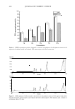
ANTI-INFLAMMATORY ACTIVITY OF C. ASIATICUM 421 PREPARATION OF THE ETHANOL EXTRACT OF CRINUM ASIATICUM The dried root of Crinum asiaticum Linne var. japonicum (0.34 kg) was extracted with 95% ethanol (1 kg) at 75°C for 3 h. Its pH was adjusted to 3.5 by adding 12 N HCl. After filtration through a 400-mesh filter cloth, the filtrate was filtered again through filter paper (Whatman, No. 5). The ethanol solution was evaporated to remove the solvent under 50°C. Lyophilization over one day yielded a yellowish powder (28.3 g, yield 8.32%). ANALYSIS OF LYCORINE CONTENT BY HPLC The ethanol extract powder of Crinum asiaticum Linne var. japonicum and lycorine · HCl (Sigma, used as a standard) were dissolved in 50% methanol. The lycorine content was investigated and calculated by HPLC (conditions: Capcell Pak Cl8 column [ 4.6 x 250 mm, Shiseido} mobile phase, 0.05 M KH 2 PO 4 and acetonitrile (95:5) flow rate, 1.0 ml/min detector, UV A. = 290 nm injection volume, 20 µl). CELL CULTURE Cells were maintained in DMEM (Dulbecco's Modified Eagle's Medium) with 10% FBS (fetal bovine serum), 50 U/ml penicillin, and 50 µg/ml streptomycin. The medium was changed every two or three days. Cells were cultured at 3 7°C in a humidified atmosphere of 95% air and 5% CO2 . CYTOTOXICITY ASSAY Raw 264.7 was seeded into 24-well plates at a density of 8 x 10⁵ cells and cultured at 37°C in 5% CO 2 . After one day, fresh medium containing 10% serum was added to the cells, which were then treated with LPS (lipopolysaccharide, 1 µg/ml), followed by treatment with the ethanol extract of Crinum asiaticum for 24 h. Cytotoxicity was evaluated by an MTT ,(3-(4,5-dimethylthiazo-2-yl)-2,5-diphenyl tetrazolium bromide [Sigma}) assay (5). After 24 h, 100 µl of 2.5 mg/ml MTT was inserted into each well, and the plates were incubated at 3 7°C for an additional 4 h. Then, the media containing MTT was discarded and the MTT formazan product was extracted with 1 ml DMSO. The amount of formazan in the culture medium was determined by the absorbance measured at 5 70 nm by an ELISA reader. Cell viability was calculated as: Cell viability(%)=(OD57o(sam p le/OD57o(control)) X 100 where OD 57 o(sample) is the absorbance at 5 70 nm of the cells treated with the sample and OD 57 o(conrrol) is the absorbance at 5 70 nm of the negative control (non-treated cells). DETERMINATION OF NITRITE SYNTHESIS Nitrite in the media was measured by the Griess assay (6) and was used as an indicator of NO synthesis in the cells. In brief, an equal volume of the culture supernatants in the Raw cell line 264.7 and the Griess solution (1:1 mixture [v/vJ of 1 % sulfanilamide and 0.1 % N-[naphthyl} ethylenlide diamine dihydrochloride in 5% H3PO4) was added into

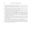
422 JOURNAL OF COSMETIC SCIENCE 96-well plates for 10 min at room temperature. The absorbance at 570 nm was measured by spectrophotometry. RNA ISOLATION AND RT-PCR OF iNOS Raw 264. 7 cells were seeded at a density of 2 x 10⁶ cells into a 100-mm dish and cultured at 37°C in 5% CO2 . After one day, fresh medium containing 10% serum was added to the cells, which were then treated with LPS (lipopolysaccharide, 1 µg/ml) and the ethanol extract of Crinum asiaticum for 8 h. Total RNA was isolated from cells with TRizol (Invitrogen) according to the instructions of the manufacturer. First-strand cDNA synthesis was performed using random hexamers. The sequences of primers are as follows: 5 '-CAGTTCTGCGCCTTTGCTCAT-3' (sense) and 5 '-GGTGGTGCGG CTGGACTTT-3' (antisense) for iNOS, and 5 '-GACGTGCCGCCTGGAGAAA-3' (sense) and 5'-GGGGGCCGAGTTGGGATGG-3' (antisense) for GAPDH. The RT PCR reaction of iNOS was reverse-transcripted at 50°C for 30 min and denatured at 96 ° C for 3 min, followed by 32 cycles at 94°C for 30 sec, 60 ° c for 30 sec, and 72°C for 1 min, and then followed by an extension step cycle at 72°C for 10 min. The RT-PCR reaction of GAPDH (glyceraldehyde-3-phosphate dehydrogenase) was reverse transcripted at 50°C for 30 min and denatured at 96°C for 3 min, followed by 32 cycles at 94°C for 30 sec, 60°C for 30 sec, and 72°C for 1 min, and then followed by an extension step cycle at 72°C for 10 min. The final products were detected with 1.5% agarose gel. The gels were photographed, and the intensity of the stained PCR fragments from photographs was quantified by densitometric analysis using Gel Doc 2000 (Bio Rad Laboratories, Segrate [Milan}, Italy). PGE2, IL-6, AND IL-8 RELEASE ASSAY Human fibroblasts were seeded at a density of 1 x 10⁵ cells into six-well plates and cultured at 37°C in 5% CO2 . After one day, fresh medium containing 10% serum was added to the cells, which were then treated with various stimuli such as H202 (5 x Hr4 M), UV (UVA 2 J/cm2 + UVB 0.2 J/cm2), SDS (sodium dodecyl sulfate), and various concentrations of sample for 48 h. The culture supernatants were used to quantify PGE2, IL-6, and IL-8 by the enzyme immunoassay kit (PGE2-Assay Design IL-6 and IL-8-Endogen) according to the protocols of the manufacturers. INHIBITION OF 13-HEXOSAMINIDASE RELEASE FROM RBL-2H3 CELLS The same procedure was followed as described previously (9). RBL-2H3 cells were resuspended in MEME (Minimum Essential Medium Eagle) media supplemented with 10% fetal bovine serum at a density of 5 x 10⁵ cells/well. Cells were dispensed into 24-well plates, and were treated with IgE (0.5 µg/ml) overnight at 37 ° C in a 5% CO2 incubator. The next morning, the cells were washed and preincubated in PIPES buffer (pH 7.2, 119 mM NaCl, 5 mM KCl, 0.4 mM MgC1 2 , 25 mM PIPES, 40 mM NaOH, 5 .6 mM glucose, 1 mM CaC1 2 , 0.1 % BSA) for 10 min at 3 7°C. The cells were treated with antigen (DNP-BSA, 1 µg/ml) for 10 min at 37 ° C. The reaction was stopped in an ice bath for 10 min, and the supernatant was used for the enzyme assay. Twenty microliters of the supernatants was added into 96-well plates and was incubated with 20
Purchased for the exclusive use of nofirst nolast (unknown)
From: SCC Media Library & Resource Center (library.scconline.org)