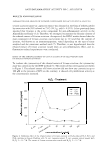
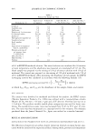
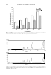
PERCUTANEOUS ABSORPTION OF OMC 389 produced by a xenon arc lamp, and unwanted spectral regions were removed by filter systems. The application amount of the tested sunscreen samples and the standard formulations was 2 mg/cm2 . The sunscreen was spread over the area with a finger stall in order to achieve a uniform film. After application, the product was allowed to dry for 15 minutes before irradiation. Air was conditioned between 20°C and 25°C during the waiting period. Each test subsite in a series was exposed to controlled amounts of simulated sunlight using the solar simulator. The MEDs (minimal erythemal doses) of the unprotected and protected skin were recorded and then the SPFs of the samples were determined (34,3 5 ). DETERMINATION OF PERCUTANEOUS ABSORPTION OF OMC USING THE STRIPPING METHOD After filing an informed consent, six healthy Caucasian volunteers (three women, three men), aged 37 ± 8 years, participated in this study. For 15 minutes they were held in a stripping room where the temperature was maintained at 20°C and the relative humidity was 50-60%. Preparations were applied onto areas of 4 cm2 , on the flexor surface of the central area of the forearm. Each assay was repeated three times. The product was applied at a dose of 2 mg/cm2 with a glass spatula, weighed before and after the deposit, and was spread uniformly over the whole area. Non-treated skin surround ing the application site was protected with adhesive tape. Protected non-occluding guards were used over the skin sites to prevent accidental product removal. Subjects wore loose long-sleeved lab coats throughout the study to minimize light exposure to the product-treated skin sites, ensuring that the sites were protected but not occluded. The Transpore tapes™ adhesive tapes were applied to the treated areas by application of a consistent pressure generated by stroking the thumb ten times along the tape (36,3 7). Stripping was performed at 0.5, 3, 5, and 7 hours after treatment under controlled conditions over 10 seconds on the same volunteers. The stratum corneum was removed by 22 tape strips with transparent adhesive tape (Transpore tape™, 3M). Strips 1-2, 3-7, 8-12, 13-17, and 18-22 were pooled separately in different groups. These strips were called group 1 (strips 1-2), group 2 (strips 3-7), group 3 (strips 8-12), group 4 (strips 13-1 7), and group 5 (strips 18-22), which introduce the different layers of the stratum corneum. Ten milliliters of methanol was added to the strips, and each sample was shaken with a vortex shaker. The amount of OMC was then determined in these solutions by HPLC (6,7 ,13,36). STATISTICAL ANALYSIS A one-way ANOV A statistical test was used to assess the significance of the differences in the concentrations of OMC in different groups of strips. In the case of a significant F value, multiple-comparison Tukey-Kramer tests was used to compare the means of the different treatment groups. Results with p 0.05 were considered to be statistically significant. RESULTS CHARACTERIZATION OF THE LIPOSOMES The liposomes made by the fusion method were morphologically homogenous multi-

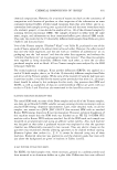
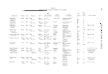
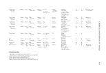
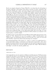
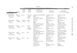
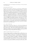
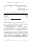
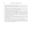
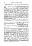
390 JOURNAL OF COSMETIC SCIENCE lamellar vesicles, as observed under optical microscope. The mean diameters of MLV liposomes containing OMC determined by PSA were 2.21 ± 0.06 µm (n = 3). The SUV liposomes were negatively stained and photographed with an electron micro scope to characterize the morphology and size distribution of the liposomes (Figure 1). These liposomes were SUV with a size of 62 ± 21 nm. In this study, liposomes with high encapsulation efficiency were prepared. Encapsulation efficiencies of MLV and SUV liposomes containing OMC were 89.66% ± 2.08 and 89.7% ± 0.7 (n = 3), respectively. Statistical analysis showed that encapsulation effi ciency of OMC is independent of liposome size and that there were no significant differences in the encapsulation efficiency of MLV and SUV liposomes (p 0.05 ). These liposomes exhibited no changes in encapsulation efficiency when stored at room tem perature for over three months. DETERMINATION OF THE SPF OF THE HOMOSALATE REFERENCE, COLIPA REFERENCE, O/W EMULSION, AND LIPOSOMES CONTAINING OMC BY IN VIVO METHOD The homosalate reference product and the COUP A high reference product were both used to validate the results of SPF determination. The SPF of the references and samples were determined by the Australian standard in vivo method. According to this method, the SPF of the homosalate standard product was calculated as 4.35 ± 0.64, and there were no significant differences between our in vivo study value (4.35 ± 0.64) and the claimed SPF (4.47 ± 1.279), with p 0.05 (Table I). The data obtained from our study show that the differences between the SPF obtained from our in vivo results (15 .2 ± 3.4) and the published SPF for COLIPA high reference (15.3 ± 2.1) were not significant. The SPF of the liposomes containing OMC was a little bit greater than that of the OMC lotion at the same concentration of OMC however, the difference was not quite signif icant. Figure 1. Transmission electron micrograph of SUV liposomes containing OMC (100,000x).
Purchased for the exclusive use of nofirst nolast (unknown)
From: SCC Media Library & Resource Center (library.scconline.org)