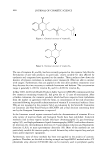
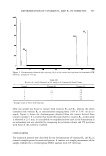
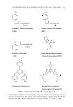
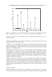
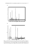
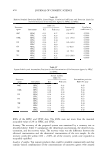
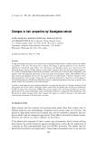
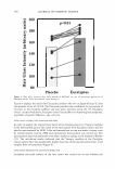
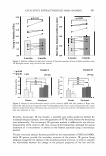
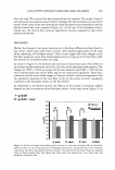
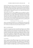
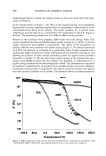
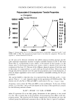
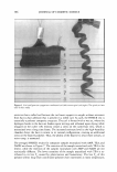
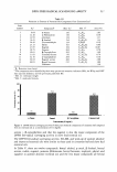
484 JOURNAL OF COSMETIC SCIENCE NANO-INDENTATION BY ATOMIC FORCE MICROSCOPE (AFM) In the case of nano-indentation by an atomic force microscope (AFM), samples of human hair fibers from Japanese females were prepared by embedding them into epoxy resin, and cutting to reveal a mirror-finished surface. The scanning of the cantilever of AFM was carried out under room temperature (25 ° C). The physical properties of the hair fiber in the cross-sectional area were obtained at the nanometer scale with a NanoScope Illa multi-mode AFM (Digital Instruments Co.), equipped with nano-indentation measure ment components. The details of the experimental technique are as reported by Ruetsch (22). Nano-indentation was carried out 25 times on the cross-sectional area of each hair fiber, sampled from two panelists. Four hair fibers per panelist were measured. Two fibers were the new-growth part of hair that was treated with the Eucalyptus extract scalp lotion, and two fibers were the new-growth part of hair that was treated with the placebo lotion. We carried out a simulation to obtain Young's modulus. The simulation model was adapted from the Hertz model (23). The AFM parameters were as follows: Cantilever: Si single crystalline probe (made by N anosensor company, NCH-1 OV, spring constant: 40 Nim) Resonant frequency: 300 kHz MICROSCOPIC METHOD IR SPECTRUM The hair section was made with a trimming knife, placed between two NaCl single crystals, and fixed. The IR spectrum was measured in the cortex region of the hair section by the microscopic method the measurement range assumed to be 6 µm2 (Figure 1). In order to obtain the proportion of the secondary protein structure of both the new-growth part of the Eucalyptus extract lotion-treated hair and the new-growth part of the placebo lotion-treated hair, the spectra of the amide I band were analyzed using the Sarver method (24). The observation condition and parameters of the microscopic IR spectrum method were as follows: • Equipped machines: Herschel FT/IR-480 Plus-type Fourier transform infrared spec- trophotometer, Irtron IRT-30-type infrared microscope QASCO) • Measuring method: Revealing, minute penetration method • Measurement parameter: Integrated times 100 times per step, resolution: 4cm - l • Detector: MCT • Aperture size: 4 x 4 µm • Mapping conditions: (a) Step: X = 10 µm and Y = 10 µm. (b) Point: X = 6 and Y = 6, 36 points in total • Hair samples: Eucalyptus-treated hair: eight hair fibers placebo hairs: eight hair fibers RESULTS USAGE TEST Figure 2 shows the results for bending measurements for sampled hairs of Group 1 after three months use of EL (Eucalyptus extract, 3%) and PL by the half-head test. The ratio

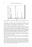
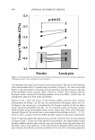
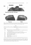
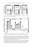
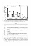
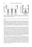
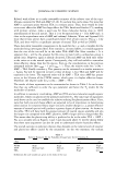
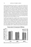
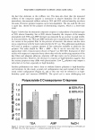
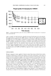
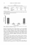
EUCALYPTUS EXTRACT-INDUCED HAIR CHANGES 485 (Thickness:about 2 J1 m) Measuring Area(6 J1 m2) Medulla Figure 1. Microscope IR measurement. The outer layer (gray) indicates the cuticle, the circle in the center indicates the medulla, and the white area between the cuticle and medulla is the cortex. The gray square means measuring area of IR by the microscopic method. of averaged Young's moduli of the hair fibers of the EL side against that of the PL side was 1.067 ± 0.041 (p = 0.0122, t-test). It can be said that the increase in hair elasticity by the use of EL was significant. Photographs of the root part of the hair on both sides of the head were taken, after natural drying and binding up of the hair of the top part of the head. Figure 3 shows photographs of the head of one panelist, as an example of the difference in appearance after EL usage. In the view from the back (a), the improvement of the stand-up at the root part of the hair can be seen, and in the view from the side (b), the visible scalp at the root of the parted hair on the EL side was noted to be narrower than that of the PL side. The results of the interviews with the panelists after three-month usage are summarized in Table I. More than 70% of the panelists had a good feeling about the change in the physical properties of the hair fibers on the EL side. As mentioned above, it has been confirmed that the panelists sufficiently recognized these changes in hair fiber proper ties. Figure 4 shows the difference in hair gloss intensity between the hair fibers treated with the Eucalyptus extract lotions and those treated with the placebo lotion for three months at the root. The results for all panelists indicate that the hair gloss intensity increased at the re-growth part by using the Eucalyptus extract scalp lotion. The total increase in hair gloss intensity was about 10%. In order to confirm the dose dependence of the Eucalyptus extract in the scalp lotion, Els containing 0.5%, 1.0%, and 3.0% Eucalyptus extract were used in the half-head tests
Purchased for the exclusive use of nofirst nolast (unknown)
From: SCC Media Library & Resource Center (library.scconline.org)