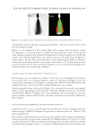
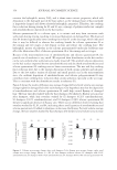
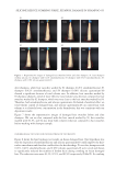
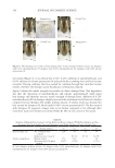
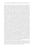
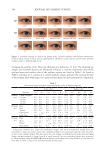
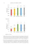
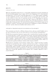
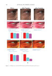
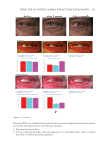
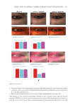
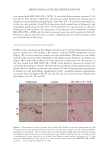
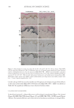
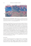
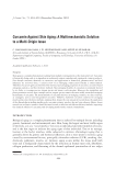
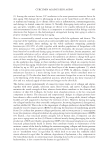
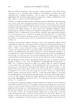
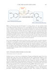
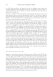
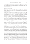
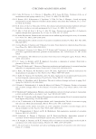
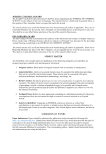
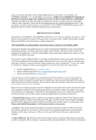
400 JOURNAL OF COSMETIC SCIENCE Elastin fibers in lesional (Figure 3F) skin were greatly decreased compared with those in nonlesional (Figure 3E) skin. The elastin fibers were torn and sparse. However, in the skin treated with MEL/NB-UVB +MTK (Figure 3H), elastin fibers were greatly increased. In the skin treated with MEL/NB-UVB +MTK, elastin fibers were increased in all cases (100%, 17/17) even in the treated skin after only 8 days (Table IC, patient 38). Elastin fibers became thick and encircled the rete ridge. However, in the skin treated with MEL/NB-UVB alone, no recovery of elastin fibers was observed (Figure 3G, Table IA). By contrast, in the skin treated with MEL/NB-UVB +MT, increase in elastin fibers was observed in 14 of 17 patients (82.4%, Table II). This difference in the recovery rate of elastin fibers between MEL/NB-UVB alone and MEL/NB-UVB +MT was statistically significant (p 0.001), whereas the difference between MEL/NB-UVB +MTK and MEL/NB-UVB +MT was not statistically significant. Moreover, in the patients treated with MEL/NB-UVB +MT (29, 33, and 34), no recovery of elastin fibers and no differentiation of melanocytes and melanoblasts was observed. Difference in the frequency of complete recovery of elastin fibers between MEL/NB-UVB +MTK (100%, 17/17) and MEL/NB-UVB +MT (42.9%, 6/14) was statistically significant (p 0.01). DISCUSSION The present study demonstrated that the loss of epidermal melanocytes and melanoblasts as well as the reduced elastin fibers in the vitiliginous skin was restored by MEL/ NB-UVB exposures and MTK irrespective of gender and age. Even in patient 49, who received only three sessions of MEL in addition to MTK, melanocyte/melanoblast differentiation was induced. However, MEL/NB-UVB alone failed to induce melanocyte/ Figure 3. Azan staining revealed well-developed collagen fibers (Coll) in the nonlesional (A), lesional (B), MEL/NB-UVB-treated (C), and MEL/NB-UVB +MTK–treated (D) skin in a similar density. By contrast, elastin fibers (Elas) were greatly decreased in the lesional (F) and MEL/NB-UVB–treated (G) skin compared with nonlesional skin (E). However, in the skin treated with MEL/NB-UVB +MTK (H), elastin fibers were dramatically increased. Scale bar =25 µm. Original magnification ×400.

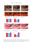
401 MESENCHYMAL STEM CELL-DERIVED FACTORS melanoblast differentiation and recovery of elastin fibers. These results suggest that MEL/ NB-UVB exposures cannot induce melanocyte/melanoblast differentiation, but they can promote melanocyte/melanoblast differentiation-inducing effects of MTK. Hachiya et al. (12) reported that stem cell factor was increased in keratinocytes after exposures of UVB (305 nm). Stem cell factor might be one of the promoting factors that can help the differentiation-stimulating effects of MTK toward melanocytes and melanoblasts. In the present study, even in the recovered epidermis that possessed a normal size of melanocyte population, epidermal melanin pigmentation was poor. Formations of melanosomes and dendrites in melanocytes as well as the transport of mature melanosomes to surrounding keratinocytes may need a long time. Epidermal melanin pigmentation seems to be greatly increased only after active and continuous transport of numerous mature melanosomes to keratinocytes. Therefore, it is reasonable to think that the skin possessing melanocytes and melanoblasts induced by the treatment with MEL/NB-UVB +MTK requires a long time to establish obvious epidermal melanin pigmentation. UCB-MSCCS is known to contain numerous growth factors such as basic fibroblast growth factor, hepatocyte growth factor, and vascular endothelial growth factor (13). These growth factors are also known to induce the proliferation and differentiation of melanocytes (14,15). The combination of these growth factors present in UCB-MSCCS may contribute to the induction of melanocyte differentiation in cooperation with a MEL/NB-UVB–derived factor such as SCF (12). Moreover, these growth factors are known to stimulate the proliferation of fibroblasts (16). Increased fibroblast proliferation may elicit an increase in elastin synthesis. It has been reported that dermal melanocytes are intimately connected with elastin fibers (17). Moreover, normal mouse melanoblasts expressed elastin-binding protein, and elastin- derived hexapeptide stimulates melanin synthesis and dendricity of melanocytes (18), suggesting that the interaction between melanoblasts and elastin-binding protein is started early in the development of mice. In normal human skin, elastin peptides in the presence of FFC also stimulated the proliferation and differentiation, DOPA reactivity, melanogenesis, and dendritogenesis of epidermal melanocytes (19). In addition to these previous reports, the present findings that elastin fibers in the dermis treated with MEL/NB-UVB +MTK were increased and reached the basement membrane of the epidermis may suggest that the interaction between elastin fibers and epidermal melanocytes/melanoblasts is indispensable for the differentiation of melanocytes in the vitiliginous skin. The present results that the recovery of elastin fibers in the treated lesions with MEL/ NB-UVB +MT compared with MEL/NB-UVB +MTK was greatly reduced may suggest that UCB-MSCCS contains upregulating factors toward elastin fiber development. It remains to be studied in a future study what substances included in UCB-MSCCS are involved in upregulating elastin fiber production. Our future study may be expected to contribute to the development of new cosmetics that are effective for treating vitiligo development. CONCLUSION MTK supplemented with UCB-MSCCS in combination with MEL/NB-UVB exposures may be one of the useful treatment modalities to recover vitiligo within a relatively short time. Taken together, our methods are expected to be useful for developing new cosmetics for vitiligo patients and new vitiligo treatments. Our treatments are thought to be permanent ones, since we observed that the melanocytes in the treated lesions remained
Purchased for the exclusive use of nofirst nolast (unknown)
From: SCC Media Library & Resource Center (library.scconline.org)