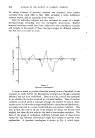
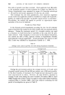
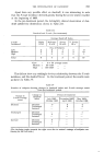
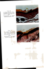
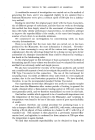
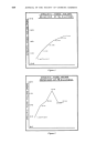
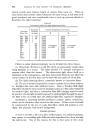
THE INVESTIGATION OF DANDRUFF Apart from any possible effect on dandruff, it was interesting to note that the P.ovale incidence declined greatly during the severe winter weather at the beginning of 1963. In the pre-treatment period, the fortnightly clinical observation of dan- druff yielded the distribution shown in Table III. Table III Dandruff and P. ovale (Pre-treatment). P. ovale Incidence* Low Medium High Average Dandruff Index Slight Moderate I Severe V. Severe 0--•5 5--•10 10-+15 15-+ 6 10 1 Nos. of Subjects 2 1 2 2 0 0 4 3 Total 6 4 17 *Low = 0-+ 50 average count Medium = 50-+150 .... High ---- 150-+320 .... This did not show any strikingly obvious relationship between the P.ovale incidence, and the dandruff level. In the treatment period, the results were as shown in Table IV. Table IV Number of subjects showing changes in dandruff index and P. ovale average count from pre-treatment period. Change in Change in Period of treatment dandruff P.ovale average index July 1962- November 1962- May 1963- count October 1962 April 1963 October 1963 -- Decrease Decrease 3 5 4 Unchanged -- 8 3 Increase -- -- -- Unchanged Decrease -- -- 1 Unchanged 5 -- -- Increase -- -- -- Increase Decrease 6 -- -- Unchanged 1 -- -- Increase -- -- -- , (The declining totals towards the right were due to natural wastage of subjects con- tinuing for the full term.)

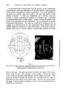
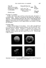
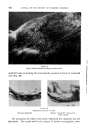
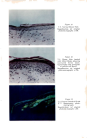
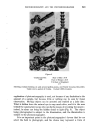
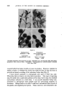
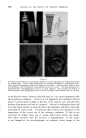
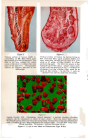
622 JOURNAL OF THE SOCIETY OF COSMETIC CHEMISTS These results showed coincident decrease in both P.ovale and dandruff in some individual cases but not in all in some cases, a fall in the dandruff index coincided with an increase in P.ovale in the early treatment period. Unfortunately, we did not manage to reduce infection virtually to nil for a prolonged interval and observe the clinical result. The findings from November 1962 to April 1963 may be regarded with suspicion owing to the abnormally cold weather. This was readily apparent in the P. ovale counts and was probably shown to some extent in the dandruff indices, in contrast to the untreated long-term series. It is also desirable to repeat the earlier suggestion that P.ovale may have an allergic rather than a toxic role if this were so, exacerbation and resolution would not necessarily be expected to coincide with infection and disinfection on a relatively short time-scale. HISTOLOGICAL STUDIES The lack of clear-cut evidence pointing to a micro-biological causation of dandruff suggested the need for histological study, which might have offered other possible mechanisms. For this purpose, it was essential to appreciate the dynamic nature of cellular activity in the epidermis and to regard the various layers seen in cross-section as representative of the changes taking place in depth. Epidermal cell-division is restricted mainly to the basal layer adjoining the dermis. In the stratum spinosum the precursors of keratin are first formed as tonofibrils and as granules of keratohyalin. The granules become more numerous and distinct as the cells move towards the surface, forming the stratum granulosum. Above this layer it is thought that the keratohyalin becomes converted to a fibrillar form in association with the tonofibrils. Orientation of the fibrils parallel to the surface may account for the bire- fringence of the next layer, the stratum lucidurn. Finally, the completely keratinized cells constitute the stratum corneum the horny cells retain little recognizable structure and have normally lost all traces of nuclear chromatin. The typical layers are easily demonstrable in the thick skin of the pall or sole but are not so clearly differentiated in the thinner epidermis of the scalp. In particular, the stratum granulosum is not continuous and may be difficult to detect in some tissue specimens. Stratum corneum normally desquamates as small, powdery particles. A Sellotape stripping of the outer skin shows that nothing but the outline of the cell shape remains. When dandruff is present, on the other hand, microscopic examination of the scales shows the presence of large amounts of nuclear chromatin. A cross-section of the scale shows that stratum corneum has been shed together with a large number of imperfectly keratin- ized cells still retaining their nuclei. This type of condition, in which the
Purchased for the exclusive use of nofirst nolast (unknown)
From: SCC Media Library & Resource Center (library.scconline.org)