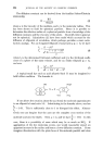
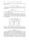
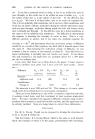
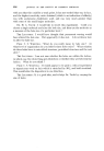
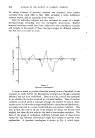
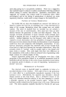
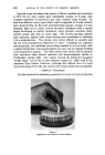
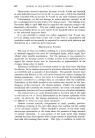
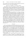
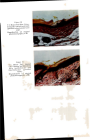
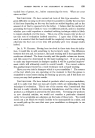
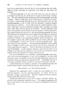
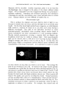
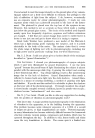
648 JOURNAL OF THE SOCIETY OF COSMETIC CHEMISTS Figure 5 Corrosion on the inside of a pressurized dispenser, lower left taken by method illustrated by dotted lines. Lower right illuminated by method indicated on continuous lines. Here grazed illumination reveals the depth of the effect. High incident light as seen here produces a lot of reflection, demonstrated to all, in colour. The incorporation of a behind-the-lens Iris-diaphragm used at a small aperture, will increase the depth of field necessary when taking shots such as the lower right. Ektachrome Type B cut film. to see that the source, whatever the bulb may be, is in correct alignment with the lamphouse condenser. If this is out of alignment the condenser will not project a pencil beam of light in the line of the optical axis, and therefore uniform illumination will not be obtained. Instead of uniformity there will be colour bands around, or along the side of the specimen, and this is especially noticeable in colour work. I would now like to draw your attention to the importance of the correct use of the substage condenser. If this is not corrected for height, blue, red or orange bands may border the image. This effect increases with the increase of magnification. If the source is not imaged at the iris-diaphragm, an enlarged colour image of the

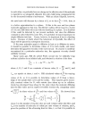
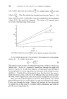
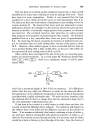
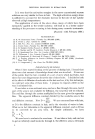
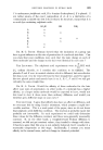
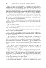
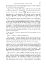
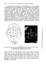
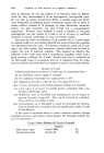
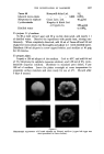
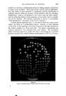
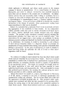
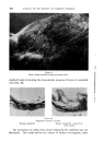
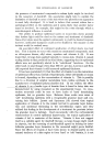
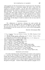
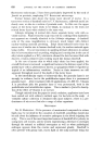
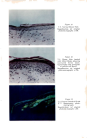
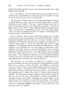
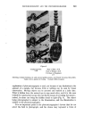
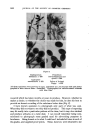
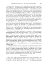
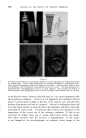
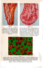
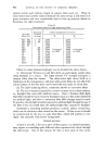
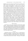
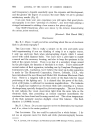
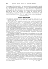
Figure 7 Vertical section of human eyelid in- corporating an 82C filter which gives the pleasant pale blue background. This filter in no way chan•es the colour values of the sta{n but colours the clear areas in the specimen. Photographed without an eyepiece attached to the microscope. Figure 8 Vertical section of human lip incorporating an 82A correction filter, again to give a better and more attractive background to that of a pure white or off white, so often revealed in colour photomicrography. The filter was placed in the light path, above the source. A large subject such as this, and that depicted in Fig. 7, is difficult to illuminate, and the extreme edges must be as sharp as the centres. As for Fig. 7, only an objective was used to make this recording. Figure .9 Aspirin Crystals X40. Illustrating "optical staining", a method whereby colourless subjects can be reproduced in colour of one's choice. Here the direct light was a Wratten 58 (green) filter providing the background, whereas the crystals were illuminated with a Wratten 29 (red) filter attached to the indirect light. Two 6V 30W lamps used. 25mm objective X4 eyepiece bellows extension 40 cm. Figures 7, 8, and .9 were made on Ektachrome Type B film.
Purchased for the exclusive use of nofirst nolast (unknown)
From: SCC Media Library & Resource Center (library.scconline.org)