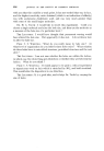
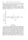
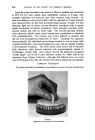
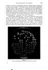
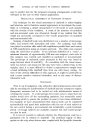
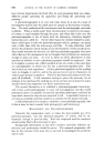
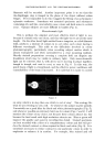
THE INVESTIGATION OF DANDRUFF 627 of the organism and the demonstration that a simple fatty ester will produce dandruff-like lesions on a laboratory animal, all tend to keep alive the concept of a microbial causation. On the other hand, parakeratosis is a relatively non-specific response to irritation and it is too early to form any firm conclusions. We have been impressed by the parallel work on Tinea Versicolor (14) which might implicate Pityrosporum orbiculare formation of antibodies to the antigen produced by this organism has been demon- strated by immunofluorescence studies. This suggests the need to consider a possible allergic type of mechanism in dandruff and, in the present state of knowledge on this subject, it would be unwise to rule out any route for further study. ACKNOWLEDGEMENTS The collaboration of numerous colleagues has made possible the publication of this investigation and acknowledgement is due especially to P. H. Cryer, H. Dixon, B. G. Overell, J. K. Herd and Miss M. Uttley. The histological work was carried out by D. B. Jones, and the photography by H. Ashmore. (Received: 31st January 1964.) (1) (2) (3) (4) (5) (6) (7) (8) (9) (lO) (11) (12) (13) (14) REFERENCES W. S. Torgerson "Theory and Methods of Scaling" 149 (!958) (Chapman & Hall Ltd., London). S. Rivolta "Parasiti Vegetali" (1873) Abst. ex R. W. Benham "Biology of Patho- genic Fungi" 63 (1947) (Chronic Botanica Co.). R. Malassez Arch. Physiol. Serie 2,1: 451 (1874) Abst. ex R. W, Benham ibid. I. Martin-Scott Brit. f. Derrnatol. 64 257 (1952). M. A. Gordon ]kIycologia 43 az4 (lUal). M. A. Gordon J. Invest. Derrnatol. 17 267 (1951). A. Tickher Brit. J. Derrnatol. ?$ 88 (1961). E. O. Butcher J. Invest. Derrnatol. 16 696 (1951). P. Flesch and S. B. Goldstone J. Inwst. Derrnatol. 18 267 (1952). D. J. Lawrence and H. A. Bern J. Invest. Derrnatol. $1 313 (1958). E. H. Mercer Keratin and Keratinization (1961) (Pergamon Press Ltd., London). P. Flesch Proc. Sci. Sect. Toilet Goods Assoc. $? 15 (1962). A. Jarrett and R. I. Spearman Brit. J. Derrnatol. 71 268 (1959). T. H. Sternberg and F. M. Keddie A.M.A. Arch. Derrnatol. 84 999 (1961). Introduction by the lecturer It is rather surprising to find so little definite information on the causation of a universal and seemingly innocent condition such as dandruff, but chronic skin disorders in general are difficult to fathom. I hope that the paper has given some hints on the directions along which the answers may be found, and I would like to show a series of slides to supplement the informa- tion given in the paper. Several of these slides represent examples of U.V.

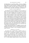
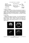
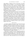
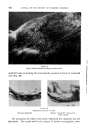
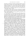
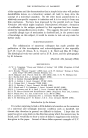
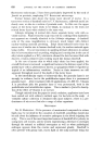
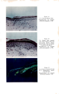
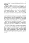
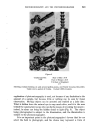
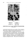
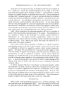
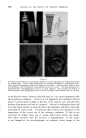
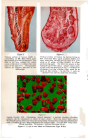
628 JOURNAL OF THE SOCIETY OF COSMETIC CHEMISTS fluorescence microscopy I have been particularly impressed by the work of Jarrett on psoriasis, employing U.V. fluorescent microscopy. Normal human skin shows the horny layer devoid of nuclei. In a transverse section of dandruff scale by U.V. fluorescence, yellowish nuclei are clearly seen, as also in a section of psoriatic scale. In this case the upper region of the horny layer tends to be bluish, in contrast to the brick red normal horny layer when stained in the same way. Sellotape stripping of normal skin shows separate horny cells with no visible nucleus. Flakes from the scalp removed by combing when dandruff is not apparent are virtually identical to the Sellotape strippings. A dandruff scale at the same magnification is obviously much larger, with distinct nuclear-staining material persisting. Ethyl oleate-induced scales from the mouse are of similar size to human dandruff scale, the nuclear material again being visible. It is not uncommon in applying irritant substances to animal skin to have reactions such as peeling, weeping and crusting but in the case of ethyl oleate applied as a 70% solution in ethanol the hair becomes filled with discrete, creamy-coloured scales looking much like human dandruff. In the case of mouse skin to which ethyl oleate has been applied, the overall thickness is increased a significant feature is the distance apart of the prickle layer cells (a phenomenon known as spongiosis) which is regarded as typical of an inflammatory condition. Nuclei or their remnants are still apparent throughout most of the depth of the horny layer. In the interfollicular region of mouse-tail skin, the granular layer is not strongly in evidence, but in the perifollicular region there is a pronounced granular layer. After treatment with 12 applications of 1,000 I.U. Vitamin A in petroleum jelly the granular layer is very pronounced, both in the perifollicular and interfollicular regions. This is similar to Jarrett's descrip- tion of the effect of Vitamin A on rat tail. Though omitted from the paper to avoid confusion, applicator tests have been carried out with ordinary nutrient agar, for bacteria on the scalp. A veritable culture collection is obtained with most subjects there is a pre- dominance of micrococci but also a range of other organisms. DISCUSSION DR. R. M^RECH^L: If the presence of unsaturated compounds in sebum lipids may be involved in the causation of dandruff, something should also be said about the influence of excessive androgens in the blood irrigating the scalp. This is one of the most important sources of dandruff after puberty in young men and women, and in women experiencing a decrease in oestrogen through the natural or artificial menopause as well as when pathological androgen appears from the ovaries and suprarenal glands. Dandruff is often present with greasy hair, excess lipid on the scalp and hair being due
Purchased for the exclusive use of nofirst nolast (unknown)
From: SCC Media Library & Resource Center (library.scconline.org)