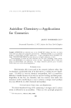
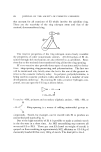
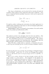
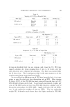
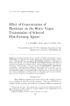
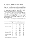
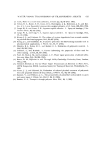
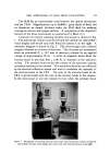
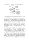
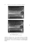
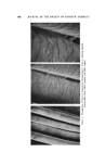
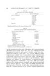
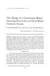
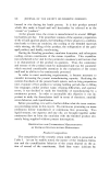
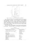
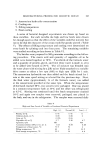
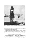
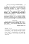
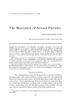
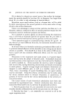
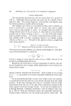
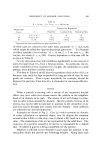
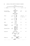
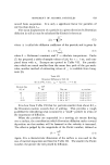
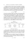
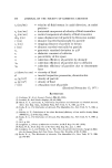
NEW APPROACHES '!'O HAIR SPRAY EVALUATION 619 The SEM fits an intermediate need between the optical microscope and the TEM. Magnifications up to 50,000X, gTeat depth of field, and no limitation on sample thickness make the SEM ideal for studying coatings on uneven and opaque surfaces. A comparison of the important features of the three instruments is summarized by Black (11). A picture of a typical scanning electron microscope is shown in Fig. I. The microscope column is on the left and the cathode ray tube (CRT) visual display and record unit with Polaroid camera is on the right. A schematic diagTam is shown in Fig. 2. The electron gun uses a heated tungsten filament as a source of electrons. The electrons are accelerated down an evacuated (2 X 10 -• mm of mercury) column by an applied voltage of approximately 25 kv. A series of magnetic lenses focus the electron beam to less than 0.01 t• (100 3,) in diameter at the specimen surface. The primary beam excites the surface of the specimen causing secondary electrons to be emitted. The secondary electrons are collected by the electron collection system and used to modulate the intensity of the electron beam in the CRT. The scan of the electron beam in the CRT is synchronized with the scan of the primary beam in the column. In the microscope at any one instant in time when the primary beam I Figure 1. Photograph of scareting electron microscope with microscope column on the left and cathode ray tube visual display and record unit with Polaroid camera on the right

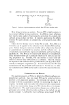
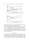
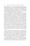
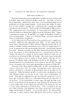
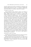
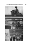
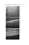
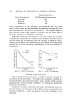
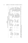
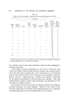
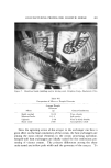
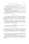
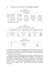
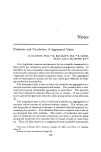
620 JOURNAL OF THE SOCIETY OF COSMETIC CHEMISTS •CO. LENS • CONDENSER I m i i LENS '-'""-' I .-,-.,-,I MAGNIFICATION SUPPLY I .' •,•/'//xm i •//x• UNIT • I LEN ...F--• I v'wl CO•LS / I SCANNING I " • •1 CIRCUITS ELECTRON- I •.•5•.•.. ' ' I I COLLECT•O, SYSTEM '""'T•--'• I,ECORD U,IT j • '•1 • ' s••l VACUUM SYSTEM / / I AMPL,F,ERS I is striking the specimen, there are secondary electrons emitted that give a corresponding spot on the CRT a certain intensity. As the primary beam moves to the next spot, the number of secondary electrons will change causing a change in the intensity of the corresponding spot on the CRT. As the "spots" are scanned, they build a black and white image on the CRT. The micrograph has a three-dimensional effect because the secondary electrons travel in curved paths. This allows electrons coming from cracks and holes of very rough surfaces to be de- tected. The degree of magnification is the ratio of the display area on the CRT to the area scanned on the specimen surface. To change mag- nification, the area scanned is increased or decreased. A decrease in magnification does not require refocusing after focusing at a higher magnification. Specimen preparation is relatively simple. The only requirement is that it must be a good electrical conductor so that a charge is not built up by the electron beam. For this reason, nonconductive materials are "prepared" hy placing them in a vacuum evaporator (3 •( l0 a mm of mercury) for deposition of a very thin coating of a conductive metal. The scanning electron microscope is an invaluable tool for studying the coatability of hair spray films on individual or small groups of hair fibers. Excellent photographs of hair, skin, and tooth surfaces can be found in Swift's article (12). Coatability can be correlated qualitatively to other important properties of hair sprays such as luster, appearance, and flaking. Generally, the better the coatability, the more natural the luster and appearance and the less flaking in the hair after combing. Coatability is a function of the properties of the hair spray resin. How- ever, where coatability is poor, the addition of certain surfactants im-
Purchased for the exclusive use of nofirst nolast (unknown)
From: SCC Media Library & Resource Center (library.scconline.org)