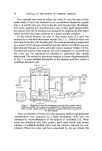
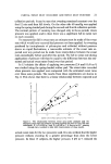
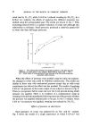
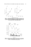
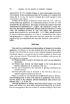
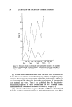
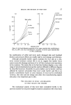
J. $oc. Cosmet. Chem. 24 15-29 (1973) ¸ 1973 Society of Cosmetic Chemists of Great Britain The use of partial sweat duct ß ß ß occlusion the elucdauon of sweat duct function in health and disease C. E. JOHNSON and S. SHUSTER* Presented on the 11th April 1972 in Oxford, at the Symposium on 'Skin Environmental responses and protection', organized by the Society of Cosmetic Chemists of Great Britain. Synopsis--A brief summary is made of the current knowledge of SWEAT GLAND FUNC- TION including the relationship between structure and function. Understanding of the role of the sweat DUCT is at present limited to speculation and circumstantial evidence. In order to characterize the function of the duct more clearly we have used our TECHNIQUE of partial sweat DUCT OCCLUSION. When pressure is applied to the SKIN surface during sweating there is a decrease in the output of sweat water, Na + , K + and urea. This change is related to the nature and magnitude of the PRESSURE and to the sweat rate. The evidence is that partial duct occlusion produced by an external pressure increases re-absorption of these substances from a distal part of the sweat duct. These findings necessarily infer the existence of a second more proximal absorptive site in the duct. Water re-absorption occurs more rapidly in the former and electrolyte re-absorption in the latter. The results suggest that the sweat duct absorbs a constant fraction of the sweat presented to it by the coil. INTRODUCTION Eccrine sweat glands are found almost exclusively in higher primates they are especially well-developed in man. These glands are found in all skin sites, although their density is not uniform. Their thermoregulatory *University Department of Dermatology, Wellcome Laboratories for Research into Skin Disease, Royal Victoria Infirmary, Newcastle-upon-Tyne. 15

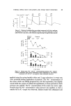
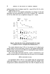
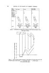
16 JOURNAL OF THE SOCIETY OF COSMETIC CHEMISTS function is of course vital to normal function particularly in hot environ- ments and during physical exertion. The sweat gland consists of a glandular tube, coiled tightly upon itself at its origin deep in the dermis, which then ascends to the epidermis through which it spirals to open onto the skin surface. Studies by three authors, Ellis (1), Munger (2) and Hibbs (3) have shown that the coiled segment is made up of cells whose structure indicates a secretory function. The structure of the cells in the straight or ductal segment is different they appear to be metabolically very active but were thought not to be secretory cells and these authors could only speculate on their function. The secretory function of the coiled segment has been characterized by Schulz, Ullrich, Fr6mter, Holzgreve, Frick and Hege (4). They inserted micropipettes into the coil and withdrew samples of sweat which were found to be isotonic with blood and their NaC1 concentration was identical to that of blood plasma and extracellular fluid. Slegers (5) and Dobson (6), using indirect methods to assess the function of the coil, also considered that it elaborated an isotonic fluid. The fact that sweat collected at the skin surface is hypotonic and con- tains low concentrations of NaC1 indicates that modification of sweat secreted in the coil has occurred, presumably in the sweat duct. Schulz et al (4) measured the electrical potentials within the sweat duct and found them to be negative with respect to the surrounding tissue. Local injection of an inhibitor to Na + transport (G-strophantin) reduced this negative potential and the sweat collected at the skin surface now had an increased NaC1 concentration. They concluded that NaC1 was normally re-absorbed in the duct and this process was responsible for the hypotonicity of sweat. Evidence has also been accumulating that the sweat duct may absorb water as well as sodium. Thompson (7) examined buried skin grafts for via- bility of the sweat glands. He showed that some glands eventually communi- cated with the surface and functioned normally. Buried glands appeared to be fully functional too, with no rupture of the sweat glands indicating that if secretion occurred total re-absorption of sweat also took place. The urea concentration in sweat is always above that of blood plasma and Schwartz, Thaysen and Dole (8) presented indirect evidence that this was due to re-absorption of water in the sweat duct. This explanation has been challenged by Brusilow (9) who provided equally good evidence that re-absorption of water did not account for the higher values of urea in sweat. More direct evidence for the re-absorption of sweat in the duct has been provided by several authors. Recently, Fasciolo, Totel and Johnson (10)
Purchased for the exclusive use of nofirst nolast (unknown)
From: SCC Media Library & Resource Center (library.scconline.org)