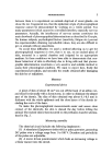
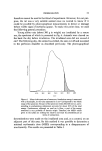
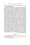
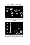
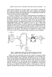
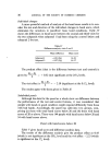
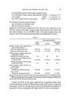
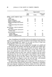
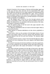
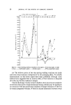
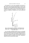
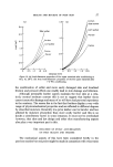
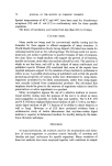
PHOREOGRAPHY 5 between them is to experiment on animals deprived of sweat glands--we chose the rat. It appeared too, that the epidermic origin of phoreographical response cannot be demonstrated by working on a whole animal. Too many parameters (which cannot be controlled) interfere, especially nervous parameters. Actually, the interference of nervous system constitutes the main drawback of phoreographical determinations as described by Kryspin. On human subjects, psychological factors (emotivity, fatigue etc.) prevent the experimentalist obtaining reproducible values, they are also difficult to get on animals without anaesthesia. To avoid these difficulties we used a method allowing us to get the phoreographical responses in vitro, that is to say, on an excised piece of skin, mounted in a special chamber and irrigated by an appropriate medium. In these conditions we have been able to demonstrate that non- linear behaviour of skin is effectively due to living cells and that phoreo- graphic determination constitutes a very sensitive and suitable method to assess their physiological condition. We want to report here, firstly the experimental procedure, and secondly the results obtained after damaging the skin by uv radiations. METHOD Experimental device A piece of skin of about 20 cm 2 was cut off the back of an albino rat, and sliced horizontally with a keratotome, in order to eliminate the deeper part of the dermis. The slice of skin obtained was about 0.1-0.3 mm in thickness with the whole epidermis and the close layers of the dermis in- cluding the roots of the hairs. To make the phoreographical measurements easier and assure dose contact of the electrode, the skin is shaved the day before killing the animal. The excised skin is then mounted on the perfusion chamber schema- tized in Fig. 2. Measuring assembly The electrical circuit includes the following implements: (1) A stimulator (Equipements industriels) or pulse generator, generating DC pulses over a voltage range from 1 to 100 V. Duration and periodicity of the pulses are adjustable. (2) A motor driven potentiometer which feeds an increasing voltage in a linear manner into the circuit.

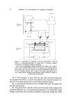
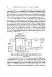
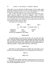
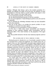
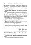
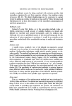
6 JOURNAL OF THE SOCIETY OF COSMETIC CHEMISTS Recorder Measuring assembly Perfusion cell I I I ,, I I 9- • •f"•'••--• I I I L_ I Figure 2. 1, Stimulator 2, voltmeter 3, driving potentiometer 4, 3M • resistor 5, measuring electrodes 6, excised skin 7, perfusion medium 8, thermostat 9, perfusion medium oxygenation. Up to now we used Parker medium, so-called '199' medium. This medium which is a very sophisticated one is due to J. F. Morgan, H. J. Morton & R. C. Parker [Proc. Soc. Exp. Biol. Med. 73, 1 (1950)] and supplied by Service des Virus de l'Institut Pasteur, Paris. It seems from recent experiments that a more simple medium (perhaps simple Ringer) could be effective enough to maintain the skin potential. (3) A 3M oe-• resistor, in series with the skin. Skin resistance being low as compared with 3M f•, it can be estimated that the intensity remains con- stant for the whole duration of a pulse (ramp-intensity clamp). (4) An electrometer (Keithley) and a recorder (Servogor) to register the transient phenomena. (5) Two silver/silver chloride electrodes (Beckman) which measure the potential difference by a conventional arrangement of saturated KC1 agar bridges constructed from polythene capillary tubing. One bridge lies inside
Purchased for the exclusive use of nofirst nolast (unknown)
From: SCC Media Library & Resource Center (library.scconline.org)