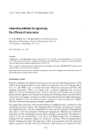
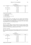
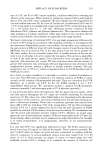
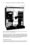
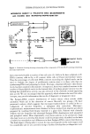
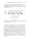
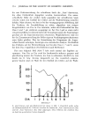
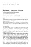
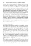
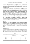
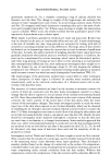
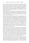
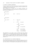
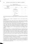
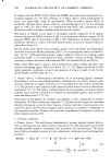
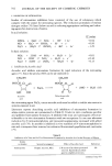
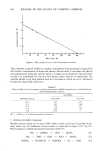
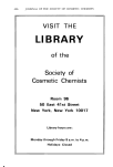
DERMATOLOGICAL INVESTIGATIONS 541 INTEGRATED DENSITY & PROJECTED AREA MEASUREMENTS with ¾1CKERS M85 MICROSPECTROPHOTOMETER monochromltor h] • sc.nner m.sk photomultip specimen SPECIMEN VIEWING SYSTEM MASKING SYSTEM flying-- spot lister scIn photomultiplier ,Integr.ted Density -- signal cted Aree Figure 4. Schematic drawing showing relationship of the components of Vickers M-85 scanning-integrating microspectrophometer been extremely fruitful in studies of the cell cycle (2). Cells in G• have a diploid or 2C DNA contents, cells in G2, a 4C content, while cells in S have intermediate values. Thus the percentage of cells with DNA contents exceeding the diploid mode can be used to evaluate the degree of proliferative activity since it is these cells that are synthesizing DNA and preparing to divide. Psoriasis, a hyperproliferative skin disease (4-6), has been studied in this manner. As expected, there was a marked increase in the number of hyperdiploid nuclei in the lesional skin of perhaps greater interest was the finding that proliferative activity was also elevated in the clinically normal-appearing skin as well. We are encouraged that this approach, which obviates the need for radio- isotopes, may provide information that can be of diagnostic or prognostic value. The Feulgen-DNA content distributions of tumor tissues often reveals subtle anomalies which aid in the detection of cancer. Recently two groups (78) have presented evidence which suggests that microspectrophotometry is useful in the cy- todiagnosis of mycosis fungoides, a malignant skin reticulosis. Microspec- trophotometric measurements were obtained in these studies from imprint specimens prepared by touching fresh biopsy material to a glass slide. Patients with clinically definite mycosis fungoides had abnormal Feulgen-DNA distributions with aneuploid and polyploid values. More importantly, even those patients in the premycotic stage who later went on to develop this disease could be prospectively identified on the basis of subtle but nonetheless real differences in their Feulgen-DNA content distributions. The final impact of being able to screen for mycosis fungoides in the early stages could be considerable and we are currently trying to further develop this cytodiagnostic tool.

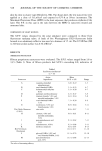
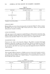
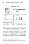
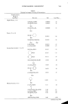
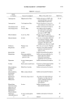
542 JOURNAL OF THE SOCIETY OF COSMETIC CHEMISTS Table I Procedures Amenable to Microspectrophotometric Measurements 1) Histochemical & Cytochemical Staining Reactions: DNA (Feulgen, Gallocyanin-chromalium, methyl green) RNA (Azure B, Pyronin Y) Histones (Alkaline Fast Green, Eosin-Fast Green) Proteins (Naphthol Yellow S, Millon, Sakaguchi) Carbohydrates (PAS) Mucopolysaccharides (Alcian Blue, Mucicarmine, Colloidal iron) 2) Enzymatic Histochemistry: Lysosomal--Bitensky Fragility Test Mitochondrial--monoamine oxidase Pentose Shunt--glucose-6 phosphate dehydrogenase 3) Redox State: Prussian blue of Chevremont-Frederic 4) Natural Pigments: Cytochrome P~450 Hemoglobin 5) Quantitative Autoradiography. Although most absorbance applications have centered on Feulgen-DNA measure- ments, the current surge of interest in microspectrophotometric analysis probably stems from recent development in other histochemical methods. Although the details of these procedures are beyond the scope of this overview, a few examples that are amenable to microspectrophotometric measurements are listed in Table I. In general, these methods enable the detection of tissue chemical changes in the picogram (10 -12 g) range with a routine accuracy of _+ 2%. The use of microspectrophotometry in conjunction with reliable histochemical methods offers several advantages over the conventional form of biochemical analyses ("grind and find"). First, it can be used to make many measurements on minimal amounts of sample tissue. Moreover, multiparameter analyses can often be achieved in the same specimen by using a combination of methods either simultaneously or sequentially. The most important advantage offered by this approach is that it allows the investigator to simply relate observed biochemical changes to the structure of the biological specimen being examined. Thus it is quite possible to measure such things as amount of mucopolysaccharides in the dermis, keratohylin content in the granular layer, the sudanophilia of lipids in sebaceous glands, lysosomal enzyme activity of the basal layer or sulfhydryl or disulfide groups of keratin in situ. With the availability of such instrumentation one no longer needs to be content with making subjective appraisals of staining intensities. Instead it is now quite easy to quantify the precise amounts of specific material of a variety of dermatological specimens from normal or diseased skin. APPLICATIONS USING AREA MEASUREMENTS The Vickers M-85 scanning-integrating microspectrophotometer allows one to measure the projected area of the specimen. We have found this facility to be extremely useful in histogeometric analyses. For example, the need frequently arises to measure the mean epidermal thickness, a parameter which is markedly influenced by disease and experimental manipulations. In the conventional approach, the value
Purchased for the exclusive use of nofirst nolast (unknown)
From: SCC Media Library & Resource Center (library.scconline.org)