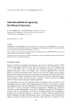
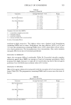
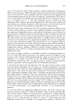
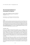
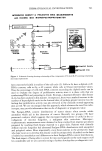
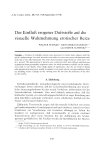
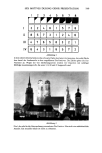
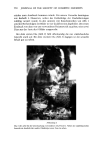
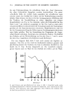
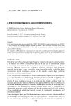
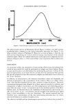
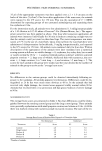
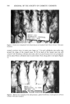
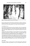
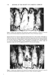
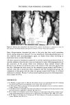
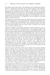
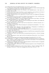
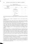
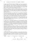
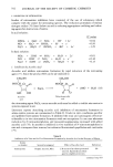
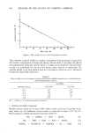
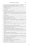
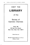
POLYMERIC FILM-FORMING SUNSCREEN 571 . ß .. .., fiR()!lP VII .q I IN I)( )W N © 3f) l•ll N. l'lXllXlliRS I C•ll PlIN. ItVL, Figure 6. Hairless mice tranquilized with Innovar©-Vet, painted with Product C, immersed in water 30 min, injected with chlorpromazine and exposed to UVL for 150 min--96 hr after irradiation Since chlorpromazine depressed the mice to the point that they would sometimes drown during immersion, Innovar©-Vet was substituted. Following administration of this relatively mild neuroleptanalgesic agent, no deaths (from either drowning or medication) were recorded thereafter. All three sunscreen formulations appeared to provide equivalent protection from ul- traviolet radiation when the mice were not immersed in water following application of the sunscreen. In the groups which were immersed, those animals treated with Products A or B were not protected due to the elution of the sunscreen agents from the skin. Although the burn scores for the immersed group treated with Product C indicate some loss of protection when compared with the nonimmersed group, the comparison of this group with the other two groups treated with Product A or B and immersed is dramatic. Product C still provided significant protection following immer- sion in water for 0.5 hr. REFERENCES (1) H. Wolska, A. Lagner and F. N. Marzulli, The hairless mouse as an experimental model for evaluating the effectiveness of sunscreen preparations, J. Soc. Cosmet. Chem., 25,639 (! 974). (2) G. Kahn and G. Wilcox, Sunscreen testing using sunlight: photographic and in vivo methods compared, J. Invest. Dermatol., 53,200 (1969). (3) T. M. Macleod and N. Frain-Bell, The study of the efficacy of some agents used for the protection of the skin from exposure to light, Brit. J. Dermatol., 84, 266 (1971). (4) M. A. Pathak, T. B. Fitzpatrick and E. Frenk, Evaluation of topical agents that prevent sunburn--supe- riority ofpara-aminobenzoic acid and its ester in ethyl alcohol, New EnglandJ. Med., 280, 1459 (1969). (5) D. W. Owens, J. M. Knox, H. T. Hudson and D. Troll, Influence of humidity on ultraviolet injury, J. Invest. Dermatol., 64, 250 (1975).

J. Soc. Cosmet. Chem., 29, 573-580 (September 1978) In vivo measurement of transepidermal water loss B. IDSON Hoffmann-La Roche, Nutley, NJ 07110. Received September 1, 1977. Presented at Annual Scientific Meeting, Society of Cosmetic Chemists, December 1976, New York, New York. Synopsis An overview is presented of the background and principle methods for MEASURING TRANS- EPIDERMAL WATER LOSS (TWL) IN VIVO. Absolute values of TWL are a function of the particular technique and experimental conditions. TWL will vary with different skin sites and rise markedly if the skin barrier is removed or affected by pathologies. Early gravimetric methods lack sensitivity and require long testing periods as well as large areas of skin. The disadvantages have caused shifts to other techniques where absorption of water vapor is followed by a sensitive physical measurement. The majority of methods are based on determining the increase in moisture content of either a current of dried air or fixed humidity air conducted over the skin. Others have sought to avoid air flow, using changes in conductivity of inorganic crystals. Methods discussed include thermal conductance, electrohygrometry, infrared radiation, electrolysis of absorbed water vapor and calculation of vapor pressure gradient in the layer of air adjacent to the skin sur- face. The mechanism may be an additive effect of neural control of eccrine sweat gland activity and stratum corneum hydration. INTRODUCTION Water exerts a major role in all well-being but particularly in skin health to maintain its desirable soft, flexible mechanical properties (1). The lack of adequate water in the up- per layer of the skin, the stratum corneum, results in dry and chapped skin (2-6). Dermatologic and cosmetic interest has focused on techniques that generate informa- tion on the state and quantity of water in the stratum corneum, the mobility of the water and the influence exerted by components of the stratum corneum on the diffu- sion characteristics of the water (1). Water is lost through skin in two ways, eccrine sweating and transepidermal diffusion. Under severe thermal stress as much as 2 1/hr may be lost as sweat. By contrast, diffu- sional or transepidermal water loss (TWL) is a steady passive process in which water vapor diffuses from the highly hydrated underlying tissues through the avascular stratum corneum, dissolves in it and diffuses to the exterior surface where it evapo- rates. Emphasis is placed on the stratum corneum since this biologically inert membrane--due to its dense, fibrous, lipoprotein matrix--represents the principle physical barrier to the penetration of molecules through the integument (7). The mag- nitude of TWL has been widely used as a measure of the effectiveness of this barrier in dermatological disease states (8-11). In pathologies such as psoriasis, ichthyoses and 573
Purchased for the exclusive use of nofirst nolast (unknown)
From: SCC Media Library & Resource Center (library.scconline.org)