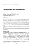
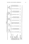
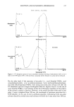
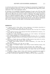
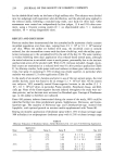
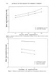
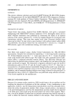
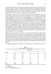
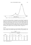
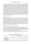
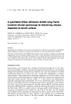
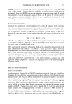
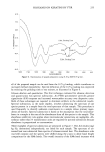
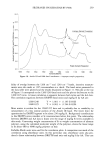
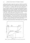
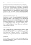
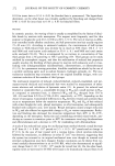
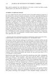
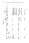
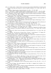
248 JOURNAL OF THE SOCIETY OF COSMETIC CHEMISTS elemental silicon and thus is non-specific for siloxanes. A siloxane-specific method was required for deposition studies. Initially, attenuated total reflectance (ATR) techniques were evaluated for hair surface analysis. The presence of siloxane was detected on amine-functional polymer treated hair. However, quantitation of the siloxane would be extremely difficult due to variability in hair/prism contact, and method development activities were discontinued. The method selected here was diffuse reflectance infrared Fourier transform spectroscopy (DRIFTS). In transmission spectroscopy, a light beam passes through a certain thickness of sample material. Energy is absorbed by the sample as a function of the molecular species present as well as its concentration. Transmission spectroscopy works well for a variety of sampling techniques such as gas, solution, mull, film, sandwich, or pellet. However, normal transmission has limitations and disadvantages for difficult samples such as solids, including fillers, powders, rubber, or cured resins. ATR is widely used for materials that are partially deformable to achieve good contact between the sample and the prism. For solid materials, such as fillers, powders, and crystalline forms, DRIFTS offers definite advantages. Generally these advantages include improved sensitivity, surface analysis, and minimal sample preparation. Diffuse reflectance involves the measurement of a spectrum from the reflected light of a sample. A perfect diffuse reflector is one which reflects light equally in all directions rather than in a well-defined path. Only a portion of this reflected light can be collected and analyzed, which leads to the requirement of a high-sensitivity spectrometer. An FTIR spectrometer with a mercury cadmium telluride (MCT) detector is well suited to meet this requirement. A diagram of the Barnes Analytical attachment used in this study is illustrated in Figure 1. m4 or sample m6 m• Figure 1. Diagram of Barnes Analytical diffuse reflectance attachment.

SILOXANES ON KERATINS BY FTIR 249 This attachment fits into the normal sample compartment of the FTIR. Light from the interferometer is reflected by the fiat mirrors, M1 and M2, onto an ellipsoidal mirror, M3. This mirror focuses the light onto the sample, which is placed in a metal cup at M4. The light is diffusely reflected from the sample, and a portion of the reflected light is collected by a second ellipsoidal mirror, M5. The light is refocused on the flat mirrors, M6 and M7, and then onto the detector. Access to the sample is from the top of the attachment, by sliding the ellipsoidal mirrors out of the way. The metal sample cup is removable for filling, and the sample cup height is adjustable for maximizing signal output. A suitable background or reference must be selected. Finely ground KBr or KCI powders are commonly used as diluents and should then be selected as the background. A review article by Griffiths and Fuller (5) provides a summary of the theory, instru- mentation required, and application examples for diffuse reflectance spectrometry. Commercial attachments for these techniques became available in 1979. Since that time, a variety of applications have been investigated. No reference in the literature was found for the use of DRIFTS with keratin fibers. In addition, the majority of papers contained qualitative information on spectral changes rather than quantitative data. A literature search concerning use of infrared spectroscopy for the analysis of keratin fibers resulted in few articles. Those papers available are generally concerned with the study of oxidized keratins showing band shifts and generation of new bands character- istic of the process. Two examples of the early work are Weston (6) and Alter and Bit-Alkhas (7) all use KBr pellet techniques. Baddiel (8) reports the use of multiple internal reflectance to study the surface structure of human hair directly rather than by grinding to form a KBr pellet. Using this technique, he shows interesting shifts in protein bands of the cuticle layer as compared to the internal cortical material. Low and Severdia (9) report the use of FTIR-photothermal beam deflection spectroscopy to char- acterize the spectrum of a single human hair in a non-destructive manner. Most re- cently, Strassburger and Breuer (10) used FTIR with a high-pressure diamond anvil cell to quantitatively measure oxidation damage to human hair. They present data on disul- fide bond cleavage by bleaching and thioglycollate waving as well as generation of sulfonate and thiosulfonate groups. The objective of the effort documented in this paper was the generation of a quantita- tive, siloxane-specific, surface analysis technique for the detection of siloxanes on hair fibers. Quantitative methods for siloxane detection are required to aid in the under- standing of the mechanism of siloxane fluid deposition. EXPERIMENTAL MATERIALS The KBr powder was purchased from Barnes Analytical. No attempt was made to keep the powder anhydrous. The KBr was ground for one minute prior to use as a back- ground reference for DRIFTS. Virgin European, natural brown hair from DeMeo Brothers, Inc. was purchased. The silicones used were trimethylsilylamodimethicones, amine-functional siloxanes, as il- lustrated in Figure 2, and derivatives of the amine functionality. The polymer con-
Purchased for the exclusive use of nofirst nolast (unknown)
From: SCC Media Library & Resource Center (library.scconline.org)