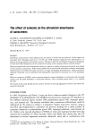
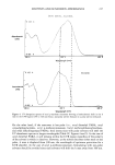
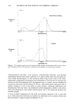
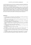
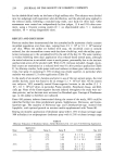
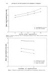
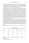
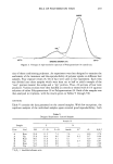
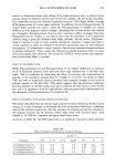
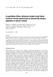
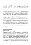
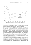
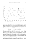
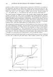
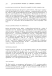
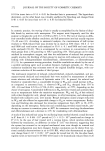
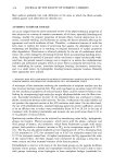
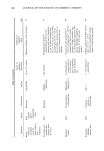
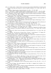
250 JOURNAL OF THE SOCIETY OF COSMETIC CHEMISTS CH 3- Si - 0 i - I OH3 LcH3 Si - Si - OH 3 I OH 3 y NH CH2-CH2-NH 2 Figure 2. Trimethylsilylamodimethicone (CTFA name). rained 100 siloxane repeating units (degree of polymerization, DP) with a 2 mole % amine functionality. The silicones were applied to the hair as 0.25 wt% diluted emul- sions. A 200-g treatment bath was used for each 12-g tress. Each hair tress was allowed to dry naturally before test clippings were collected. The entire tress length (30 cm) was used in preparing samples to provide a uniform, representative sample. INSTRUMENTATION Spectra were obtained from a Nicolet Model SX spectrometer. The spectrometer is equipped with an air-bearing Michelson interferometer, a water-cooled high-intensity globar source, and a liquid nitrogen-cooled mercury cadmium telluride detector. Spectra were collected at 2 cm-• resolution and with co-addition of 300 one-second scans. The diffuse reflectance attachment was purchased from Barnes Analytical, Stamford, Conn. The accessory is called the "Collector" Model 0030-003 and mounts in the sample compartment of the FTIR. The WIG-L-BUG grinder/mixer Model 3110B was also purchased from Barnes Analytical. The WIG-L-BUG is a vial-and-pestle device that reciprocates at 3200 rpm and is commonly used by spectroscopists for solid sam- pling and by dentists for preparation of amalgams. The sample is placed in a (2.5 cm x 1.3 cm) stainless steel vial that includes a 0.6-cm diameter stainless steel ball. TEST PROCEDURE Prepare the FTIR for operation and set-up the diffuse reflectance accessory. Grind about 0.3-0.5 g KBr powder for one minute in the WIG-L-BUG. Fill the DRIFTS macro- cup with KBr powder, level, and place in the accessory. Purge the FTIR for 5-10 minutes, maximize the signal output, and collect 300 scans as background. Repeat as necessary to eliminate CO2 and water vapor contributions. Cut hair fibers to a length of 0.25-0.50 cm. Use solvent-washed scissors and clean paper to prevent sample contam- ination. Weigh 0.20 - 0.005 g KBr powder (non-ground) and 0.05 - 0.005g cut hair fibers into a clean WIG-L-BUG vial containing the stainless steel ball. Clean the vial by rinsing with distilled water, wipe dry with a towel, rinse with methylene chlo- ride, and blow dry. Grind the KBr/hair mixture for one minute. Add all of this mixture to the DRIFTS macro-cup with the aid of a clean metal spatula. Place the sample in the

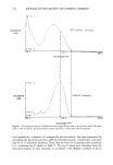
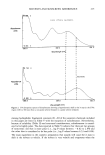
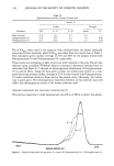
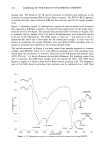
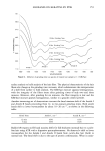
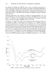
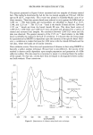
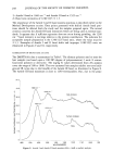
SILOXANES ON KERATINS BY FTIR 251 DRIFTS accessory, purge for 5-10 minutes, maximize signal output, and collect 300 scans for the sample. Repeat collection until CO2 and water vapor contributions are minimized. Clean the DRIFTS macro-cup and repeat preparation steps until all samples are completed. Run all samples in duplicate. Scan the IR spectra for Amide I and II band maxima. Amide I should be 1660 ___ 5 cm -1 and Amide II should be 1520 cm-1. Repeat sample if outside these limits. QUANTITATION PROCEDURE Important for quantitation was development of a linear IR response with concentra- tion. For transmission measurements, the y-scale is in absorbance units to provide a linear response with concentration. For diffuse reflectance the proper y-scale is a func- tion of reflectance. Kubelka and Munk (11) developed a general theory for diffuse re- flectance of scattering layers within powdered samples and derived the following equa- tion: fiR) = (1 - R)2/2R = k/s where R is the reflectance at infinite depth, k is the molar absorption coefficient, and s is a scattering coefficient. Software in the Nicolet FTIR system allows for easy conver- sion of the data to Kubelka-Munk units. After conversion of the spectra to Kubelka-Munk units, prepare for measurement of the band intensities of the 1260 cm -•, 1240 cm -•, and 1225 cm -• bands. Individual programs (MACROs) within the Nicolet system were developed to automatically calcu- late the band intensities with respect to a specific baseline. The baseline used for this area of the spectrum was 1359- 1178 cm- •. These programs simulate standard IR cal- culation procedures and eliminate hand calculations. Calculate the band ratios of 1260/1240, 1260/1225, and 1240/1225. If the 1240/1225 ratio is greater than 1.30, repeat the sample preparation. Calculate the mg/kg Si level of the sample using the calibration curve. RESULTS AND DISCUSSION METHOD DEVELOPMENT Sample preparation. Essentially no DRIFTS signal output results from neat hair fibers. Therefore, the non-reflective hair surface must be made reflective with the aid of a good diffuse reflector. KBr is a good diffuse reflector with no absorption bands in the infrared and was selected as the reflecting medium. However, it is necessary to produce intimate contact between the reflective and non-reflective materials for a successful DRIFTS experiment. The ideal sample preparation for surface analysis would produce a thin coating of KBr on each hair fiber. Several approaches were evaluated for mixing the hair samples with KBr. The hair fibers were cut to a length of 0.25-0.50 cm using solvent- cleaned scissors. The mixing approaches included: 1) agitation of KBr and hair in a glass vial using a wrist-action shaker, 2) grinding of the two materials in a close-toler- ance glass container, tissue grinder, and 3) grinding and mixing of materials in a Barnes Analytical WIG-L-BUG. An infrared spectrum was collected using DRIFTS after each
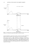
Purchased for the exclusive use of nofirst nolast (unknown)
From: SCC Media Library & Resource Center (library.scconline.org)