22 JOURNAL OF THE SOCIETY OF COSMETIC CHEMISTS cludes a large UVA component (1). An SPF-15 oxybenzone-containing sunscreen was tested in hairless mice by Bissett et al. (3) using a low-irradiance black light UVA lamp (peak emission -355 nm). These workers report no protection by this sunscreen against UVA-induced skin changes such as sagging and increased GAGs. However, avobenzone (Parsol 1789: 4-tert. butyl -4'-methoxydibenzoylmethane: peak absorption -360 nm) in a 7% solution in carbitol resulted in retardation of UVA-induced skin sagging, thickening, and amelioration of histological alterations. Harrison et al. (9) examined a 0.75 % concentration of avobenzone in an oil-and-water emulsion in hairless mice irradiated with sub-erythemal doses of UVA as emitted by unfiltered Philips TL 10 tubes (peak emission -360 nm). Unlike Bissett et al. (3), they found no protection against histologic dermal damage. This may be due, among other factors, to the low concentration of avobenzone and the small, but energetically significant, emission by these tubes of UVB at 302 and 313 nm. Broad-spectrum sunscreens have not been examined by using pure UVA with an emis- sion spectrum that closely approximates solar UVA for efficacy against photoaging changes. Thus, there is still some question regarding their efficacy against UVA I (340 nm). We decided to use the hairless mouse to compare the protective effect of two commercially available broad-spectrum sunscreens against chronic exposure to this portion of the UVA waveband. Both sunscreens had an SPF of 15, with one containing avobenzone as the UVA absorber, and the other, oxybenzone. MATERIALS AND METHODS ANIMALS AND TREATMENT GROUPS There were four groups of 12 Skh-hairless-1 female albino mice, ages 6--8 weeks (Tem- ple University Health Sciences Center, Philadelphia, PA). They were housed individ- ually under General Electric F40 GO gold fluorescent tubes, which emit no UV radi- ation (12-hour on/off cycle). Treatment groups were as follows: 1) UVA only 2) UVA with avobenzone-containing (3%) commercial sunscreen (SS-A) 3) UVA with oxyben- zone-containing (3%) commercial sunscreen (SS-B) SPF-15* and 4) unirradiated, un- treated controls. Sunscreens were applied to the entire dorsal surface of the mice (2 •l/cm 2) five minutes prior to each exposure. Vehicles for the sunscreens were not deemed necessary at the start of the experiment, nor were they available to us. The results with SS-B were totally unexpected. RADIATION SOURCE AND EXPOSURE SCHEDULE The high-intensity UVASUN 3000 lamp provided UVA I radiation (340-400 nm). At mouse level, a distance of 65 cm from the lamp, irradiance was --15 mW/cm 2 as measured with an IL 700 A research radiometer (International Light, Inc., Newbury- port, MA). The UVA sensor has peak sensitivity at -360 nm. The emission spectrum of this lamp has been published previously (2). To ensure that no radiation below 340 * The formulation tested is no longer commercially available.

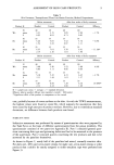
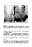
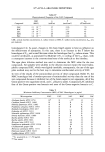
UVA SUNSCREEN 23 nm was transmitted, new filters were installed at the beginning of the experiment and again after 400 hours of use. Spectroradiometric readings of our lamp have shown that lower wavelengths begin to appear at 500 hours (unpublished results). Ambient tem- perature at the level of the mice was kept at --27øC by two small fans. An erythemic dose in naive animals with this UVA source is 50-60 J/cm 2. The exposures of 100 J/cm 2 were therefore reached gradually by 25 J/cm 2 increments over the first two weeks, to avoid erythema in the unprotected mice. With a thrice weekly exposure (M, W, F) for --32 weeks, the total cumulative dose was 8000 J/cm 2. MICROSCOPY On the Monday following the final Friday exposure, dorsal skin biopsies (2 x ¾2 cm) were taken and processed for light microscopy. The histochemical stains were hema- toxylin and eosin (H & E) for general histology, Luna's aldehyde fuchsin for elastic fibers (10), Van Gieson's for collagen, and Mowry's colloidal iron for GAGs. Slides were evaluated in a coded manner. Adjacent dorsal skin was also processed for electron microscopy. ULTRASTRUCTURE Specimens were fixed in Karnovsky's solution, followed by 1% osmium tetroxide, and were embedded in Epon 812. Ultrathin sections (70 nm) were stained with a tannic acid-uranyl acetate solution (11) followed by Reynold's lead citrate, each for 10 minutes. These were examined in a Hitachi model H700 electron microscope. COLLAGEN FIBER MEASUREMENTS Collagen fiber diameters were measured on 3-5 electron micrographs per treatment group at 42,500 x magnification. Two to three squares measuring 5 x 5 cm were drawn on each micrograph in areas where fibers were clearly in cross section. All fibers within the squares were measured. RESULTS CLINICAL APPEARANCE At the end of the 32-week irradiation period, unprotected UVA-exposed mice had the thickened, yellowed, sagging skin typically induced by this waveband (3). The skin of most SS-A-protected mice was only slightly thickened and pale, but was not distinctly yellow. A few of the mice retained the pinkish skin of unirradiated controls. SS-B- protected mice developed some scaling less than halfway through the experiment. This was judged to be irritation due to the sunscreen. By the end of the irradiation period, more than half of the animals had thickened leathery patches on the central dorsum (Figure 1). Two mice in the unprotected group had a total of three 2-mm papillomas. In the SS-B group, one mouse had one 2-rnm papilloma. There were no tumors in the unirradiated or SS-A groups.
Purchased for the exclusive use of nofirst nolast (unknown)
From: SCC Media Library & Resource Center (library.scconline.org)