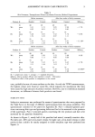
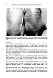
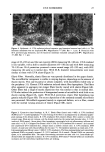
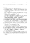
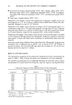
24 JOURNAL OF THE SOCIETY OF COSMETIC CHEMISTS Figure 1. UVA-irradiated mice (32 weeks: 8,000 J/cm2). Skin of SS-A-protected animal (left) has normal- appearing, pale pink skin. SS-B-treated animal (right) is scaling and leathery, with pin-point erosions (x2). HISTOLOGY General (H&oe). Unirradiated hairless mice (age "•40 weeks) have a thin 3-4-cell-layer epidermis. The collagen is mainly concentrated in the upper dermis, whereas the lower dermis contains mainly a single row of keratinizing cysts, typical for this mouse, along with a variable amount of lipocytes (1). After 8000 J/cm 2 of long-wavelength UVA, the unprotected skin was more than twice as thick, with an acanthotic epidermis of '-•7 cell layers. There was mild cellular disorder and a few foci of parakeratosis, but very little atypia. Enlarged dermal cysts proliferated up to three rows (Figure 2A). Skin protected with SS-A was mainly unthickened (Figure 2B). The epidermis was slightly acanthotic ("•5 cell layers) but was without loss of order, atypia, or parakera- tosis. Dermal cysts, while usually in a single row, were occasionally in two rows. This is within the normal range of variability for these mice. Skin protected with SS-B appeared as damaged as UVA-irradiated, unprotected skin. Epidermal atypia was more pronounced, and inflammatory infiltrates were present in the dermis and epidermis (Figure 2C). Cyst proliferation was more variable than in UVA only. Elastic fibers. The elastic fibers in unirradiated mice are thin and very sparse (12). After UVA irradiation, most specimens showed mild elastic fiber hyperplasia and a few foci of moderate hyperplasia. Fibers were also slightly thickened and tortuous (Figure 3A). With SS-A protection, the elastic fibers were as thin and sparse as in unirradiated skin

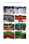
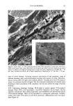
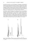
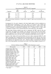
UVA SUNSCREEN 25 Figure 2. Full-thickness skin (H & E X 115). A. UVA-irradiated (8,000 J/cm 2) skin is thickened, mainly because of cyst proliferation, but the epider- mal hyperplasia contributes. The granulomatous re- action (*) that normally results from rupture of cysts is exacerbated. B. UVA-irradiated, SS-A-treated skin remains thin, with a slightly hyperplastic epi- dermis. The number of cysts is within the normal range. C. UVA-irradiated, SSB-treated skin is greatly thickened and severely inflamed. The acan- thotic epidermis shows signs of dyshesion. (Figure 3B). Specimens from the SS-B group had a degree of elastic fiber hyperplasia that was more severe than in unprotected, irradiated skin (Figure 3C). In areas of severe inflammation, elastotic clumps were beginning to form. Collagen. Normal collagen stains a bright crimson with Van Gieson's stain. Confirming earlier studies (1,2), chronic UVA irradiation did not alter the staining properties. Normal staining was also maintained in SS-A-protected skin (Figure 3D). In the SS-B specimens, there were occasional foci with reduced staining (Figure 3E), an indication of alteration to the collagen. Glycosaminoglycans. Normal hairless mouse skin contains minute amounts of the blue- staining material identified as GAGs by histochemistry (12). After UVA irradiation, dermal GAGs were increased, and the normally red-staining collagen had a blue tone, as if coated with GAGs. Intercellular epidermal GAGs were also increased (Figure 3F). SS-A-protected skin appeared similar to unirradiated skin (Figure 3G). To the contrary, SS-B specimens showed varying degrees of increased GAGs, ranging from a few foci to many. Epidermal GAGs were increased in all SS-B specimens (Figure 3H). ULTRASTRUCTURE Epidermis. In normal, unirradiated skin, basal epidermal cells are closely adherent to one another. Chronic UVA radiation induced gaps between the cells that SS-A prevented (Figures 4A & B). The basement membrane at the dermal-epidermal junction was not duplicated in either case. Collagen. Unlike with UVB (6), collagen fibers in cross section retained sharp outlines after chronic UVA exposure. The diameter of fibers in normal skin is diverse, with a
Purchased for the exclusive use of nofirst nolast (unknown)
From: SCC Media Library & Resource Center (library.scconline.org)