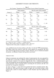
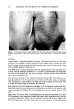
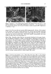
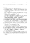
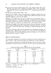
28 JOURNAL OF THE SOCIETY OF COSMETIC CHEMISTS 400 350 3OO • 2•o. o •" 200, E • 150, Z 100, 50 i Unirrad iated 12oo UVA 2 3 4 5 6 7 8 9 10 Fiber Diameter 1000 800 600 400 200 2 3 4 5 6 Fiber Diameter •6o SS-A •oo SS-B 140 350 120 300 100 ß 250 80 '• 200 6o E 15o z 40 lOO 20 50 o o 2 3 4 5 6 7 8 9 lO 2 3 4 5 6 7 8 9 Fiber Diameter Fiber Diameter Figure 5. Collagen fiber diameter. Unirradiated skin had a broad range of diameters, with the majority measuring 4-6 mm at )42,500 magnification. UVA-irradiated skin had a more narrow size range, with most fibers measuring 3-5 mm at )42,500 magnification. With SS-A protection, fiber diameters were similar to those in unirradiated skin, whereas with SS-B, fiber diameters resembled those in unprotected, UVA-irradiated skin. VASCULATURE The cutaneous vasculature appeared to be extremely sensitive to UVA-induced injury. The basement membrane surrounding most vessels was greatly duplicated, up to 14 layers, compared to the normal 3-5 (Figure 7A). In addition, endothelial cells showed

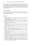
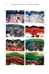
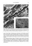
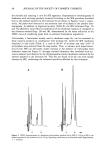
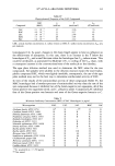
UVA SUNSCREEN 29 Figure 6. Elastic fibers. A. UVA radiation produced elastic fiber hyperplasia in mouse skin, which nor- mally contains sparse elastic tissue. Many fibers were elongated and densely coated with black-staining elastin matrix (--)). Masses of microfibrils (*) lacking elastin coating were present. Magnification X 20,800. Bar = . 05 Ixm. Inset: Uncoated microfibrils (•r) at higher magnification. Magnification X 37,500. Bar = .05 l•m. signs of severe damage, including extensive vesiculation of the cytoplasm, areas of extreme thinning, gaps, and mitochondrial swelling with rupture of cristae. Mitochon- dria in various other cell types were similarly affected. Virtually all of the UVA-induced injury was prevented by SS-A (Figure 7B). An occasional endothelial cell showed some thinning or mild vesiculation of the cytoplasm, but this was within the range of normal findings. SS-B. Confirming histologic findings, SS-B failed to protect against UVA-induced damage. Elastic fibers were hyperplastic, microfibril deposition was increased, vascular basement membranes were greatly duplicated, and endothelial cells showed the typical UVA-induced damage. These will be described in a subsequent publication. Notably, unlike with either UVA alone or with SS-A protection, inflammatory cells were abun- dant (Figure 8).
Purchased for the exclusive use of nofirst nolast (unknown)
From: SCC Media Library & Resource Center (library.scconline.org)