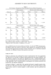
26 JOURNAL OF THE SOCIETY OF COSMETIC CHEMISTS

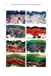
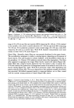
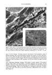
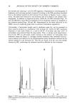
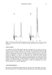
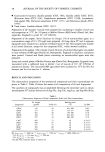
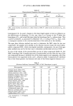
UVA SUNSCREEN 27 Figure 4. Epidermis. A. UVA radiation-induced extensive gap formation between basal cells (--). The basement membrane was not duplicated (11•). Magnification X7,800. Bar = I Ixm. B. Protection with SS-A prevented gap formation, and the basement membrane remained unduplicated (11•). Magnification X7,800. Bar = 1 Ixm. range of 45-245 nm and the vast majority (80%) measuring 90-140 nm. UVA rendered it less variable, with a shift to smaller diameters (45-150 nm) and with 80% measuring 70-120 nm. SS-A protection preserved a more normal range (45-220 nm), with 80% measuring the same as in normal skin. With SS-B, diameter measurements were more similar to those with UVA alone (Figure 5). Elastic Fibers. Normally, elastic fibers are very sparsely distributed in the upper dermis. The microfibrillar component is visible to varying degrees, depending on the amount of elastin matrix. This can be partial or almost complete, with microfibrils visible only at the periphery (11). Chronic UVA radiation induced elastic fiber hyperplasia. The fibers often appeared to aggregate into longer fibers heavily coated with elastin (Figure 6A). Other fibers had a range of matrix deposition that was similar to that in normal skin. UVA also induced the production of disorganized masses of micro fibrils with little or no elastin coating (Figure 6A, inset). With SS-A protection, elastic fiber hyperplasia was greatly reduced (Figure 6B). The excess deposition of the randomly arrayed microfibrils was prevented. Microfibrils appeared mainly in organized fashion, as in a fiber, coated with the normal varying amounts of elastin (Figure 6B), inset). Figure 3. Connective tissue histology. A, B, C. Elastic fibers (Luna's stain X460). A. UVA irradiated (8,000 J/cm2)•A mild hyperplasia (--)) and twisting of the fibers is accompanied by an abnormal purple- pink staining of the dermal matrix. B. UVA irradiated, SS-A treated--Elastic fiber content is within the normal range, fibers are not tortuous, and the dermal matrix has the normal pale-lavender color. C. UVA irradiated, SS-B treated--Severe elastic fiber hyperplasia intermingled with an inflammatory infiltrate and mast cells (--)). D, E. Collagen (Van Gieson's stain x 370): D. UVA irradiated SS-A treated--As in normal and unprotected UVA irradiated skin, collagen stains a uniform crimson color. E. UVA irradiated, SS-B treated--A small area with reduced affinity for the stain (--). The dyshesive epidermis is clearly seen. F-H. Glycosaminoglycans (Mowry's stain X 460): F. UVA irradiated--The blue-staining dermal and intercellular epidermal GAGs are greatly increased. G. UVA irradiated, SS-A treated--Dermal GAGs are extremely sparse, as in unirradiated skin. The major blue-staining structures are mast cells (--). Intercellular epi- dermal GAGs are marginally increased. H. UVA irradiated, SS-B treated--Dermal GAGs are moderately increased, and the intercellular epidermal GAGs are striking.
Purchased for the exclusive use of nofirst nolast (unknown)
From: SCC Media Library & Resource Center (library.scconline.org)