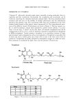
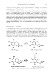
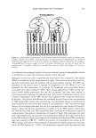
SKIN DELIVERY OF VITAMIN E 257 eases, phototoxicity, photosensitivity, and skin aging. In cutaneous tissues, solar UV irradiation induces immediate damage, leading to erythema and sunburn, and is gen- erally believed to be responsible for the delayed damage leading to premature aging and cancer. Electron spin resonance (ESR) spectroscopy has been used to confirm irrefutably that UV exposure leads to the generation of a host of free radical species by as yet uncharacterized mechanisms. Topical administration of antioxidants is one approach to diminishing oxidative injury (37). Shindo eta/. (33), while studying the antioxidant defense mechanism in murine epidermis and dermis, found that o•-tocopherol levels decreased with irradiation in both layers to approximately the same degree. Vitamin E is the major lipophilic antioxidant of exogenous origin found in tissues and is an obvious choice for enhancement of antioxidant protection by topical application. A variety of antioxidants, especially vita- min E and ubiquinol, are present in skin. Topical application provides an efficient means of enriching the tissue in protective antioxidants such as vitamin E. The cosmetic industry's main claim for vitamin E has been as a "natural moisturizer" to relieve dry skin and indirectly to aid in concealment of wrinkles and facial lines perceived as characteristics of aging and dry skin. However, studies carried on for more than a decade have now revealed significant benefits of vitamin E beyond moisturization of dry skin. Thiele eta/. (38) used vitamin E concentration as a marker of oxidative damage and found that ozone depleted topically applied but not inherent vitamin E, suggesting that ozone probably attacks the outermost layers of the skin, where the vitamin E presumably is the most concentrated. Ozone exposure (10 ppm for 2 h) caused lipid peroxidation in cutaneous tissues, and prior vitamin E application was found to ameliorate this damage. ESTERS OF (x-TOCOPHEROL o•-Tocopherol functions as an antioxidant when the phenolic hydroxyl of its chromanol ring is free (unesterified) (39). The free -OH can function as a scavenger of free radicals or singlet oxygen, usually being itself oxidized in the process to the semiquinone or quinone. The hydroxyl group can be protected from oxidation by esterification with the carboxyl group of an organic acid, forming esters such as the acetate or succinate derivatives, which are thought to reduce the antioxidant activity of the tocopheryl acetate or succinate esters to zero. The esterified forms of o•-tocopherol are stable to oxidation on storage. o•-Tocopheryl acetate, a liquid oil, when applied to the skin acts as a prodrug and has to release the active o•-tocopherol by a suitable hydrolysis reaction. o•-Tocopheryl acetate, being a lipid, is thought to diffuse across cell membranes, enter cells, and be associated with other hydrophobic membranous structures present in the cells such as the mitochondria and nuclear membranes. Kamimura and Matsuzawa (40,41) have suggested two routes for absorption of o•-tocopherol through the skin: (a) from the stratum corneum into the epidermis and then the dermis, and (b) through the hair follicles, by way of the pilosebaceous canal and into outer root sheaths and even- tually into the dermal tissue. In their study they did not show that the acetate ester was hydrolyzed to the free form, though they have speculated that it may occur. Tocopheryl linoleate is believed to be a superior moisturizing agent, with both the tocopherol and linoleic acid playing their respective roles. However, in studies using linoleate, the difficulty in distinguishing between the effect of tocopherol from the fatty acid should be borne in mind.

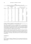
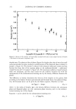
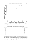
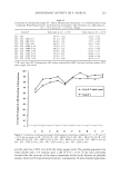
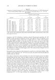
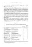
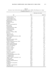
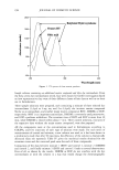
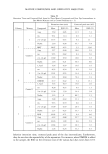
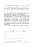
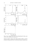
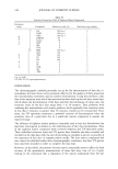
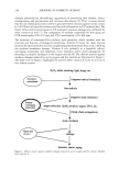
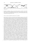
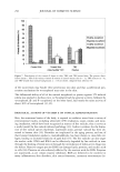
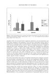
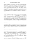
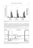
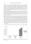
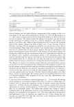
258 JOURNAL OF COSMETIC SCIENCE PHOTOPROTECTION Acute solar-simulated UV irradiation can induce oxidative damage in skin by over- whelming the skin's antioxidative capacity. Two of the early cellular events following exposure to UV light are the induction of lipid peroxidation (42) and suppression and replicarive DNA synthesis due to DNA damage (43,44). Both of these appear to be related to UV-induced free radical generation within the cells (45). (x-Tocopherol has been shown to be effective in protecting against the carcinogenic and mutagenic activity of ionizing radiation and chemical agents (46). While oral supplementation of (x-to- copherol failed to increase its concentration in the skin (47), topical application of vitamin E homologues strongly increased vitamin E levels in skin of hairless mice (38,30). Lopez-Torres eta/. (48) studied the effect of (x-tocopherol on the antioxidant network in UV-induced oxidatively damaged murine skin. They hypothesized that topical (x-to- copherol supplementation modulated the antioxidant network in the skin and bolstered its antioxidant capacity. The four principal antioxidant enzymes--superoxide dismutase (SOD), catalase (CAT), glutathione peroxidase (GPx), and glutathione reductase (GR)• were studied along with lipophilic antioxidant (x-tocopherol and hydrophilic antioxi- dants ascorbate and glutathione in murine epidermis and dermis. (x-Tocopherol was topically administered (5 mg/cm 2) along the back and sides of female hairless mice 24 hours before irradiation. Irradiation with a solar simulator at 290-400 nm was carried out after the mice were anaesthetized. The epidermis and dermis were gently separated with a scalpel after heating at 55øC for 30 sec. Lipid hydroperoxide levels, a sensitive marker of oxidative damage, were measured in the epidermis and dermis. Topical (x-tocopherol application resulted in a 62-fold increase in the epidermal and a 22-fold increase in the dermal concentration of (x-tocopherol. Table I gives the concen- trations of vitamin E in murine epidermis and dermis after topical application and UV irradiation. Even after an acute UV irradiation, the concentrations were still higher than control levels in both layers. Levels of lipid hydroperoxide increased in both the epi- dermis (11-fold) and dermis (threefold) after UV irradiation. Results are given in Figure 8. In the epidermis, (x-tocopherol treatment resulted in a significant reduction of this increase in lipid hydroperoxides. However, such protection was not afforded in the dermis. One reason for this could be the differences in tissue composition and antioxi- dant distribution. Twenty-four hours after topical application of (x-tocopherol, SOD activity was increased in the dermis, CAT activity was decreased, and GR activity remained unchanged. Table I Concentrations of Vitamin E in Murine Epidermis and Dermis After Topical Application and UV Irradiation Control UV-irradiated Vitamin E Vitamin E + Tissue (nmol/g skin) (nmol/g skin) (nmol/g skin) UV (nmol/g skin) Epidermis 16.5 + 4.4 (4) 10.0 + 3.4 (4) 1025.6 + 140.9 (5) 198.2 + 29.7 (4) Dermis 5.6 + 0.94 (4) 3.7 + 0.69 (4) 123.8 + 21.7 (5) 57.4 + 13.9 (5) Data are mean + SE. Number of animals in parentheses. Adapted from reference 48.
Purchased for the exclusive use of nofirst nolast (unknown)
From: SCC Media Library & Resource Center (library.scconline.org)