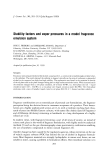
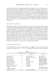
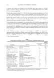
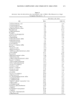
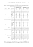
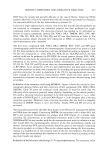
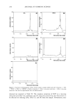
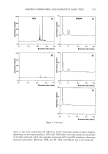
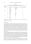
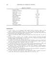
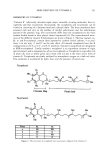
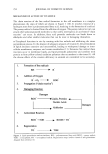
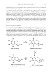
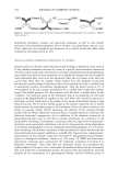
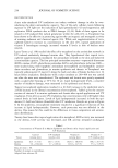
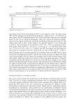
SKIN DELIVERY OF VITAMIN E 271 solution, 38.6% of the applied dose was recovered in the viable epidermis and dermis the amount detected in the horny layer was 7.12%, and the residual fraction persisting on the surface was 54.3% of the applied dose (109). I, vivo stzzdy. To establish the extent to which ot-tocopheryl acetate was bioconverted to ot-tocopherol, Beijersbergen van Henegouwen eta/. (96) studied the i, vivo permeation of vitamin E and its prodrug across rat skin membrane. Both 2.5% ot-tocopherol and 2.5% ot-tocopheryl acetate solutions in ethanol were applied to specified areas on the back of rats. At various time intervals the animals were sacrificed and the dorsal skin removed. The epidermis was separated from the dermis using 2 M KI for 1 h at room temperature. The epidermis was then removed with a scalpel. The authors found that both ot-tocopherol and its acetate behave similarly with regard to the penetration and horizontal migration through the epidermis. The pH of the stratum corneum is estimated to be about 5.0, and that of the viable tissue is about 7.4. The aromatic hydroxyl group in ot-tocopherol (pKa about 10, comparable to various phenol derivatives with similar molecular structures) is not dissociated. Hence the difference between ot-tocopheryl acetate and ot-tocopherol with regard to its physico- chemical parameters that determine skin transport was negligible. After a period of 5 h after a single application of ot-tocopheryl acetate, the amount of ot-tocopherol found did not differ significantly from that already present. Even after five days the amount of ot-tocopherol found in the epidermis did not differ significantly from that already found, indicating only minor amounts of hydrolysis. After correction for the ot-tocopherol already present without application of o•-tocopherol, hydrolysis amounted to less than 1% of the ot-tocopherol in the stratum corneum and about 5 % in the viable layer after five days. It was concluded that vitamin E acetate can be considered a prodrug that very slowly releases minute amounts of the protector vitamin E. A limitation of this study was that it did not take into consideration the whole skin, i.e., the epidermis and the dermis. The enzymatic activity in the dermis, with its system of capillary blood vessels, would be higher than the epidermis, and this may result in the higher production of ot-tocopherol from the acetate. Alberts et aL (110) conducted a Phase II cancer prevention study i, vivo to evaluate whether topically applied ot-tocopheryl acetate was absorbed in human skin and me- tabolized to the free or other forms. A cream containing ot-tocopheryl acetate (125 mg/g) was rubbed on the forearms of eleven subjects twice daily for three months. Punch biopsies were taken at the beginning and end of the time period. Results (Table IV) showed an elevated level of ot-tocopheryl acetate after three months of topical cream administration. However, the cleavage capacity of human skin was extremely low or absent, and free ot-tocopherol concentrations ranged from 38.9 + 17.9 lng/g skin at baseline to 36.3 + 20.9 lag/g skin after three months of topically applied ot-tocopheryl acetate cream. I, vivo experiments offer the advantage that skin viability is constantly maintained but can be frustrated by the difficulties of differentiating between skin metabolism and systemic metabolism. EFFECT OF FORMUL^T•O•S EmMsio,s. Emulsions are preferable to simple solutions because of their universal solu-

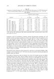
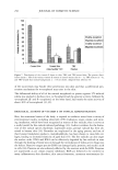
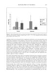
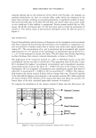
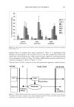
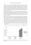
272 JOURNAL OF COSMETIC SCIENCE Table IV Skin Concentrations of tx-Tocopherol Acetate, Free tx-Tocopherol, and y-Tocopherol From Skin Biopsies of Subjects Treated for Three Months With Topically Administered tx-Tocopheryl Acetate Cream Skin concentrations (lag/g) Sample Baseline biopsy 3-Month biopsy o•-Tocopherol acetate 5.9 -+ 11.8 256.3 a _+ 195.5 tx-Tocopherol 38.9 -+ 17.9 36.3 b _+ 20.9 y-Tocopherol 6.0 _+ 3.9 4.4 c -+ 2.3 Statistical significance: a P = 0.068 b p = 0.465 c P = 0.273 vs. corresponding baseline values. Adapted from reference 110. bilizing behavior and the higher effective concentration of the vitamins in the oil or water phase for the same initial added amount. Forster et al. (101) and Kietzmann et (111) have used an isolated perfused bovine udder (BUS) model as a substitute for an in vivo test on humans. The authors studied the influence of emulsion type and structure on the penetration of vitamins E and A as cosmetic ingredients. The vitamins were applied at an infinite dose. Adhesive tape stripping was used to remove the outermost layers of the stratum corneum (ten layers of horny cells, 10 pm). Sections 20 pm in thickness, extending 200 pm through the epidermis into the top layer of the dermis, were dermatomed. Three times more vitamin E was absorbed from a w/o cream than from the oil solution from the same amount applied. This has been attributed to the greater thermodynamic activity of vitamin E in the emulsion. In multiphase systems, the distribution coefficient and the concentration of the active ingredient are dependent on the effective active ingredient concentration present in the solubilizing phase. The vitamin E absorbed into the top skin layer from the two o/w emulsions was greater than that absorbed from the oil solution, but less than that from the w/o emulsion. The authors found the stratum comeurn to be a strong penetration barrier against vitamin E. The influence of vitamin E on stratum corneum hydration and water-binding capacity was tested in o/w and w/o emulsions. The composition of the emulsions is given (112). For the stratum corneum hydration study the emulsions were applied on the forearm by volunteers and the hydration was tested by capacitance measurements. A combined in vitro/in vivo test was carried out to study the water-binding capacity. The test prepara- tions were applied evenly over the sole of the foot. Stratum corneum samples were taken using a stratum corneum scraper. This stratum corneum was completely hydrated in vitro in a humidity chamber (100% humidity, H20) over seven days. Measurements were made with an ultrabalance. In the case of the o/w emulsion, repeated application (n -- 15) increased the stratum corneum hydration, and vitamin E enhanced this effect. In contrast to vehicle alone, 5 % vitamin E led to a statistically significant increase of stratum corneum water-binding capacity (P = 0.0209). For the w/o emulsion, a 5% vitamin E content led to the best enhancement of stratum corneum hydration. In both types of emulsions the hydrating effect led to comparable results. The authors have assumed that vitamin E exerts a stabilizing effect on the bilaminate structure of the epidermal barrier lipids, which is thought to be responsible for the hydration (113). This study suffers from the limitation that application of the o/w emulsion was carried out for 14 days, whereas the w/o emulsion was applied for only eight days, and hence a strict comparison between both types of emulsions cannot be made.
Purchased for the exclusive use of nofirst nolast (unknown)
From: SCC Media Library & Resource Center (library.scconline.org)