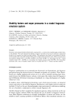
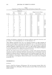
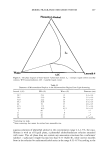
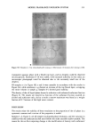
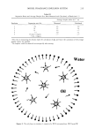
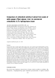
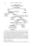
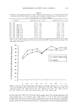
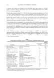
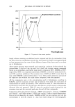
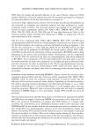
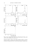
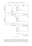
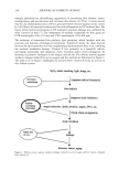
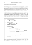
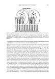
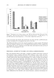
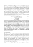
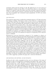
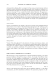
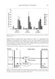
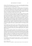
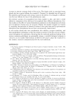
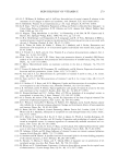
SKIN DELIVERY OF VITAMIN E 265 mulation depend also on the interaction of the vehicle with the skin--for example, an emulsion formulation can have an occlusive effect under which the hydration of the horny layer increases, resulting in increased penetration. A significant number of meta- bolic processes take place in the skin (95) and these can only be taken into account under in vitro conditions if skin viability is maintained. Various animal models like rat (96), mouse (30,38,48), and human skin (35,40) have been used to study the permeation of vitamin E. The various stages of percutaneous absorption across the skin are given in Figure 9. SKIN DISTRIBUTION Topical bioavailability and the kinetics of absorption of the tocopherols and tocotrienols have been evaluated by determining the localization of ot-tocopherol in comparison to or- and •/-tocotrienol in hairless mouse skin at various time points after topical adminis- tration (97). The concentrations of or- and •/-tocotrienol and 0t-tocopherol after topical administration of a 5% solution of the homologues in polyethylene glycol-400 for 0.5, 1, 2, or 4 h was measured in murine skin layers. The skin layers were divided depending on their thickness from stratum corneum downwards, as shown in Table II. The application of 5% vitamin E resulted in a 200- to 2000-fold increase in the skin ot-tocopherol content over that of control mice. The uppermost layer of the skin (5 pm, SC1) contained the highest •/-tocotrienol, ot-tocotrienol, and ot-tocopherol concentra- tions (pmol/cm2/p) of any of the layers (P 0.0001) for each of the vitamin E forms. Furthermore, SC1 ot-tocopherol concentrations were significantly greater than those of either SC1 •/-tocotrienol (P 0.0001) or SC1 ot-tocotrienol (P 0.0001). The relation- ship between the various vitamin E forms did not change with time. Vitamin E applied to the skin had the highest concentrations in the stratum corneum (SC1) when expressed per micron of skin. If the thickness of the various skin layers was taken into account, the lowest layers of the skin contained appreciable amounts of vitamin E. To compare the distribution of the various vitamin E forms into the skin layers, the I II I I II I II I i .• I I • I I • I I • I I I ," Stratum Comeum I II I I ! I I II I [ I I I I I II I I I I I I'• I I I I I I II I I1• I II I I II I ! ß ! ! I N. Pene•tion- Pertcation l•,o•ption •' Epidermi Basal Membrane Dermis Blood Vessel Figure 9. Stages of percutaneous absorption. Adapted from reference 101.

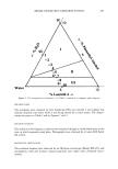
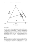
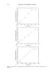
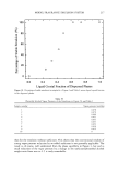
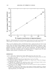
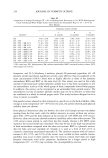
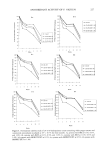
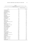
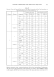
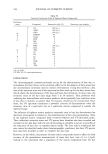
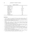
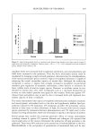
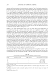
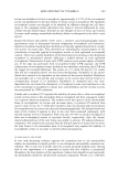
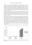
266 JOURNAL OF COSMETIC SCIENCE Table II Designation of Skin Thickness of Cryosections Using a Tape-Stripping Procedure Designation Layer Thickness (pm) SC 1 Stratum corneum 5 SC2 Stratum corneum 5 E 1 Epidermis 10 E 2 Epidermis 10 PD Papillary dermis 100 D Dermis 400 SF Subcutaneous fat =100 Adapted from reference 97. percentage of each form was expressed relative to its respective total. The lower layers (PD and D) contained the major portion of applied vitamin E due to the thickness of these layers. The PD represents about 16% of the total skin thickness, but obtained about 40-50% of the vitamin E homologues. This layer contains the sebaceous glands- lipid secretory organs, which may account for the affinity of this layer for vitamin E. The dermal layer had 30-40% of the vitamin. SF contained about 11-14% of the skin vitamin E. The percentage of •-tocotrienol in SC1 (10 + 5%) was significantly (P 0.01) greater than in SC2 (2 _+ 1%), E1 (2 + 2%) or E2 (2 _+ 2%) and lesser than in PD (40 _+ 15%) or D (36 + 15%, P 0.0001). The SC1 percentage of o•-tocotrienol or percentage of o•-tocopherol was also significantly less than in PD or D (P 0.0001). The percentage of o•-tocotrienol appeared to accumulate more in the PD, while a larger fraction of o•-tocopherol was found in the D. The results are shown in Figure 10. In all cases the percentage in PD of vitamin E was greater than its respective percentage in SF. The authors noted that within the first 0.5 h (first time point measured), vitamin E homologues penetrated through the entire skin to the SF layer, though it was unclear whether this rapid penetration was into skin cells (keratinocytes or fibroblasts), around the cells in the skin lipids, or down the hair follicles into the deepest layers. Trivedi et a/, (98) proposed that topically applied vitamin E functions as a penetration enhancer. The authors investigated the potential for permeation enhancement of a model semi- polar solute, hydrocortisone, across human cadaver skin in the presence of vitamin E and found an enhancement in the permeability of hydrocortisone by vitamin E. SKIN METABOLISM OF o•-TOCOPHERYL ESTERS Skin is not a passive barrier that merely restricts the diffusion of chemical agents into the body. The skin is a viable, metabolizing membrane that can metabolize an assortment of topically applied substances before they become systematically available. Much em- phasis has been spent in the last several years on examining the influence of skin metabolism on percutaneous absorption. Certain highly reactive compounds, such as benzo[a]pyrene, testosterone, and estradiol may be significantly metabolized during their percutaneous absorption. Vitamin E acetate or other esters of o•-tocopherol are often used as the prodrug forms of vitamin E due to their stability under atmospheric con- ditions. However, it is theoretically impossible for the acetate to have an effect as an antioxidant since phenolic hydroxyl of the tocol nucleus is chemically protected by the acetate esterified to it. Despite this, vitamin E acetate affords protection against the
Purchased for the exclusive use of nofirst nolast (unknown)
From: SCC Media Library & Resource Center (library.scconline.org)