HAIR PHOTOPROTECTION BY DYES 389 (7) (8) (9) (10) (11) (12) (13) (14) C. Dubief, Experiments with hair degradation, Cosmet. Toilerr., 107, 95-102 (1992). R. Beyek, G. S. Kass, and C. F. Meyer, Elasticity and tensile properties of human hair. II. Light radiation effects,J. Soc. Cosmet. Chem., 22, 667-678 (1971). E. Tolgyesi, Weathering of hair, Cosmet. Toilerr., 98, 29-33 (1983). L. Wolfram "The Reactivity of Human Hair--A Review," in Hair Research, C. E. Orfanos et•/., Eds. (Springer-Verlag, New York, 1981), pp. 479-500. P. Alexander, M. Fox, and R. F. Hudson, The reaction of oxidizing agents with wool, Biochem. J., 49, 129 (1951). J.B. Speakman, Mechano-chemical methods for use with animal fibers, J. Textile Inst,, 37T, 102 (1947). C. Pande, FT-Raman spectroscopy: Applications in hair research, J. Soc Cosmet. Chem., 45, 257-268 (1994). I.J. Miller and G.J. Smith, Protection against phototendering of wool by metal salts and mordanted dyes, J.S.D.C., 111, 103-106 (1995).

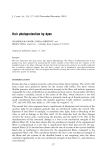
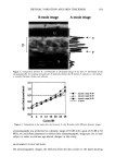
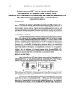
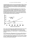
j. Cosmet. Sci., 52, 391-397 (November/December 2001) Diurnal variation affects age-related profile in skin thickness KAZUE TSUKAHARA, YOSHINORI TAKEMA, SHIGERU MORIWAKI, TSUTOMU FUJIMURA, and GENJI IMOKAWA, Biological Science Laboratories, Kao Corporation, 2606 Akabane, Ichikai, Haga, Tochigi 321-3497, Japan. Accepted for publication August 15, 2001. Synopsis We have previously demonstrated that over the course of each day there are changes in skin thickness that can be measured by B-mode ultrasonography. This suggests that there is a shift in dermal fluid from the face toward the legs by gravity, resulting in a diurnal variation in skin thickness. Therefore, age-dependent profiles in skin thickness were evaluated by B-mode ultrasonography in the morning or in the afternoon for 130 normal Japanese females aged 18-83 years. Three areas of the face (the forehead, the corners of the eye, and the cheeks) were measured as distinctively sun-exposed areas while the flexion side of the forearm was measured as a weakly sun-exposed area. A weak correlation between skin thickness and age was found in all areas measured (positive for the forehead, the corners of the eye, and the cheeks negative for forearms) in the morning but not in the afternoon, when only a weak positive correlation was observed in the cheek. These results indicate that when measuring skin thickness, an appropriate time for taking measurements should be selected with consideration of the movements of derreal fluid over the course of each day. INTRODUCTION For noninvasive measurement of skin thickness, the usefulness of ultrasonography has been reported (1) and has been extensively used to study skin thickness according to age, sex, and areas of the body (2-5). Concerning the association between skin thickness and age as evaluated by A-mode ultrasonography, skin thickness has been reported to de- crease with age in forearm skin, which is weakly exposed to the sun (6-8). We previously demonstrated an age-related increase in the skin thickness of the face, tested as a sun-exposed area by A-mode ultrasonography (9). Recently, Gniadecka and Jemec (10) also reported an age-related increase in the skin thickness of the face by B-mode ultra- sonography. A similar increase in facial skin thickness in an older group compared with a younger group has been reported (11). We have recently demonstrated a diurnal variation in skin thickness measured by B-mode ultrasonography, which suggested a Address correspondence and reprint requests to Genji Imokawa. 391
Purchased for the exclusive use of nofirst nolast (unknown)
From: SCC Media Library & Resource Center (library.scconline.org)