j. Cosmet. Sci., 52, 391-397 (November/December 2001) Diurnal variation affects age-related profile in skin thickness KAZUE TSUKAHARA, YOSHINORI TAKEMA, SHIGERU MORIWAKI, TSUTOMU FUJIMURA, and GENJI IMOKAWA, Biological Science Laboratories, Kao Corporation, 2606 Akabane, Ichikai, Haga, Tochigi 321-3497, Japan. Accepted for publication August 15, 2001. Synopsis We have previously demonstrated that over the course of each day there are changes in skin thickness that can be measured by B-mode ultrasonography. This suggests that there is a shift in dermal fluid from the face toward the legs by gravity, resulting in a diurnal variation in skin thickness. Therefore, age-dependent profiles in skin thickness were evaluated by B-mode ultrasonography in the morning or in the afternoon for 130 normal Japanese females aged 18-83 years. Three areas of the face (the forehead, the corners of the eye, and the cheeks) were measured as distinctively sun-exposed areas while the flexion side of the forearm was measured as a weakly sun-exposed area. A weak correlation between skin thickness and age was found in all areas measured (positive for the forehead, the corners of the eye, and the cheeks negative for forearms) in the morning but not in the afternoon, when only a weak positive correlation was observed in the cheek. These results indicate that when measuring skin thickness, an appropriate time for taking measurements should be selected with consideration of the movements of derreal fluid over the course of each day. INTRODUCTION For noninvasive measurement of skin thickness, the usefulness of ultrasonography has been reported (1) and has been extensively used to study skin thickness according to age, sex, and areas of the body (2-5). Concerning the association between skin thickness and age as evaluated by A-mode ultrasonography, skin thickness has been reported to de- crease with age in forearm skin, which is weakly exposed to the sun (6-8). We previously demonstrated an age-related increase in the skin thickness of the face, tested as a sun-exposed area by A-mode ultrasonography (9). Recently, Gniadecka and Jemec (10) also reported an age-related increase in the skin thickness of the face by B-mode ultra- sonography. A similar increase in facial skin thickness in an older group compared with a younger group has been reported (11). We have recently demonstrated a diurnal variation in skin thickness measured by B-mode ultrasonography, which suggested a Address correspondence and reprint requests to Genji Imokawa. 391

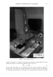
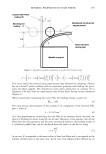
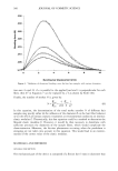
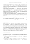
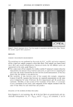
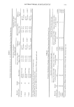
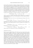
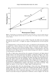
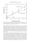
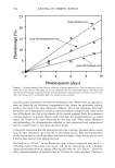
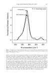
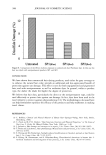
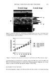
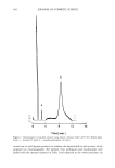
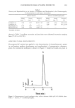
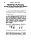
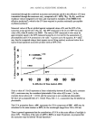
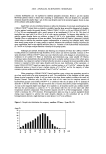
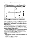
392 JOURNAL OF COSMETIC SCIENCE shift in dermal fluid from the face toward the legs by gravity over the course of each day, resulting in the elicitation of a diurnal variation in skin thickness (12). It is thus conceivable that the diurnal variation in skin thickness is also modulated by aging as a result of differing degrees of dermal fluid accumulation between the upper and lower body. To clarify the effects of diurnal variation on age-related changes in skin thickness, we have now studied skin thickness in the morning and in the afternoon by B-mode ultrasonography at three sites on the face (the forehead, the corners of the eye, and the cheek) as sun-exposed areas and on the ventral side of the forearm as a poorly sun-exposed area. Here we report that there is an age-dependent slight increase in the thickness of all the facial skin sites measured in the morning, which may be primarily ascribed to an increase in an age-related accumulation of dermal fluid in the face during a night's sleep. MATERIAL AND METHODS SUBJECTS Skin thickness was measured on 130 healthy Japanese females aged 18-83 years. All subjects had no renal or cardiac diseases. Measurements were performed on each volun- teer at three sites (the forehead, the corners of the eye [1.5 cm from the inner canthus], and the cheek [1 cm down from the top of the cheekbone]) on the face as sun-exposed areas and on the ventral side of the forearm as a poorly sun-exposed area. Before measurements, each subject washed her face with a liquid face wash (Kao Corporation, Tokyo, Japan) and stayed in a room at a constant temperature of 23øC and a relative humidity of 40% for 30 minutes for acclimation. MEASUREMENT BY B-MODE ULTRASONOGRAPHY Ultrasonography by B-mode was performed using a UX-01 ultrasonic diagnostic system (RION Co. Ltd., Tokyo, Japan) at 23øC and 40% relative humidity, as described previously (13). Briefly, the apparatus has two kinds of frequency, 15 MHz and 30 MHz, with a running width of 20 mm and 10 mm, respectively, in the direction of X, Y, and Z. The scanning mode has a B-mode with a scan time of 50 ms. The resolution ability is within 90 pm or 180 pm at 30 MHz or 15 MHz, respectively, focused at 30 mm. The velocity of ultrasound in the skin is 1530 m/sec. We measured the skin using B-mode ultrasonography under the above conditions to obtain ten ultrasonographic images per site in each subject. The B-mode ultrasonographic images obtained were printed (on thermal recording paper for Mitsubishi video copy processor SCT-K65H) using a video printer (SCT-P65 video copy processor, Mitsubishi Electric Corp, Tokyo, Japan). In preliminary experiments using A-mode ultrasonography (Dermascan A, Cortex Tech- nology, Denmark, at 1580 m/sec), we measured the skin thickness on the ventral forearms of ten females (aged 25-34 years, mean age 30 years) and compared that with the skin thickness measured using B-mode ultrasonography at the same site with dif- ferent frequencies, dynamic ranges, and gains (Figure 1). Since there was close agreement between the skin thickness measured by the above two methods (Figure 2) when B-mode
Purchased for the exclusive use of nofirst nolast (unknown)
From: SCC Media Library & Resource Center (library.scconline.org)