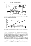
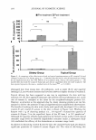
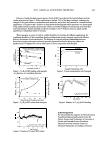
162 JOURNAL OF COSMETIC SCIENCE wounds. It is important to note that the temporal order in which the treatments were applied did not affect the results, indicating that a ten-minute rest period between applications was sufficient to produce accurate responses. REFERENCES (1) Physicians' Desk References, For Nonprescription Drugs and Dietary Supplements, 20th ed. (Medical Econom ics Company, Montvale, NJ, 2001), p. 611. (2) V. P. Shah, Progress in methodologies for evaluating bioequivalence of topical formulations, Am. J. Clin. Dermatol., 2, 275-280 (2001). (3) Y. N. Kalia, I. Alberti, A. Naik, and R.H. Guy, Assessment of topical bioavailability in vivo: The importance of stratum corneum thickness, Skin Pharmacol. Appl. Skin Physiol., 14(S1), 82-86 (2001). (4) H.J. Weigmann, J. Lademann, R. von Pelchrzim, W. Sterry, T. Hagemeister, R. Molzahn, M. Schaefer, M. Lindscheid, H. Schaefer, and V. P. Shah, Bioavailability of clobetasol propionate Quantification of drug concentrations in stratum corneum by dermatopharmacokinetics using tape stripping, Skin Phannacol. Appl. Skin Physiol., 12, 46-53 (1999). (5) L.A. Nylander-French, A tape stripping method for measuring dermal exposure to multifunctional acrylates, Ann. Occup. Hyg., 44, 645-651 (2000). (6) T. Frodin and M. Skogh, Measurements of transepidermal water loss using an evaporimeter to follow the restitution of the barrier layer of human epidermis after stripping the stratum corneum, Acta Derm. Venereol., 64, 537-540 (1984). (7) S. J. Bashir, A. L. Chew, A. Anigbogu, F. Dreher, and H. I. Maibach, Physical and physiological effects of stratum corneum tape stripping, Skin Res. Technol., 7, 40--48 (2001). (8) J. Pinnagoda, R. A. Tupker, T. Agner, and J. Serup, Guidelines for transepidermal water loss (TEWL) measurement, Contact Dennatitis, 22, 164-178 (1990). (9) B. Jones and M. G. Kenward, Design and Analysis of Cross-Over Trails (Chapman and Hall, New York, 1990), pp. 197-199.

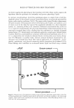
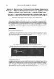
J. Cosmet. Sci.) 55, 163-176 (March/April 2004) Salivary films on hydroxyapatite studied by an in vitro system for investigating the effect of metal ions and by a quartz-crystal microbalance system for monitoring layer-by-layer film formation Y. TANIZAWA, N.JOHNA, Y. YAMAMOTO, and N. NISHIKAWA, Analytical Research Center, Lion Corporation, Hirai, Edogawa-ku, Tokyo 132-0035, Japan Accepted for publication January 26, 2004. Synopsis The salivary film or the acquired pellicle is a protein film formed initially on the enamel surface of teeth. Such a film plays an important role in enamel protection, but is also an initial substructure for the formation of plaque and the cosmetically undesirable colored stain. The composition and the structure of the film are still essentially unknown because of the difficulty of its isolation for characterization. The purpose of this study was to investigate the effect of some metal cations on the salivary film or the pellicle formation, and also to clarify the mechanism of development. First, using infrared spectroscopy (IR) and X-ray photoelec tron spectroscopy (XPS), the in situ-farmed film in the mouth was confirmed to contain selectively adsorbed well-known proteins. Then, in vitro studies have demonstrated that Ca2 + ions enhance film formation at the initial stage in virtue of Ca bridging and, interestingly, that Mg2 + ions oppositely inhibit the formation. Furthermore, the quartz-crystal microbalance (QCM), utilized successfully for the first time to study the salivary film, has shown the possibility of an alternate accumulation mechanism by which the surface charges on the film are effectively reversed by the opposite charged proteins. INTRODUCTION Extrinsic dental stains are cosmetically undesirable colored deposits (1,2) that lie on the surface of the teeth. The prevention and the removal of the stains by abrasive toothpastes have been investigated (3-6). The dental stains are often associated with culculus, plaque (7), and adsorbed salivary film materials termed the acquired enamel pellicle (8), though the mechanism of formation is still speculative. In some cases, substances like tea and coffee incorporated into the pellicle produce the staining as a result of their inherent colors. The pellicle materials are also stained as the result of aging and chemical interactions (3,9). Therefore, in the present work attention has been directed to the salivary film or pellicle. The secretion from human salivary glands contains a number of proteins or glycoproteins having a strong affinity for enamel and hydroxyapatite (HAP). These proteins selectively 163
Purchased for the exclusive use of nofirst nolast (unknown)
From: SCC Media Library & Resource Center (library.scconline.org)