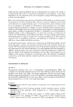
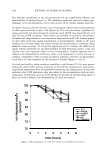
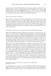
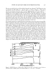
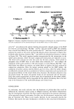
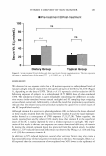
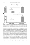
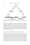
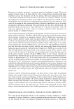
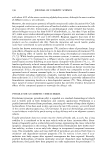
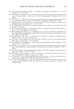
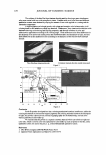
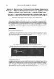
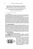
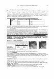
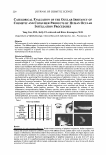
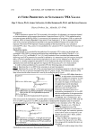
224 JOURNAL OF COSMETIC SCIENCE ADVANCED HISTOLOGICAL TECHNOLOGY AND THREE-DIMENSIONAL IMAGING: A FRUITFUL PARTNERSHIP TO VISUALIZE THE HUMAN SKIN MICRO-ANATOMY AND CHANGES IN CUTANEOUS STRUCTURES Gilles Pauly, M.D., Marie-Danielle Vazquez-Duchene, Ph.D., Dominique Gauche, Jean-Luc Contet-Adooneau, M.D., Christine Jeanmaire, Ph.D., Louis Danoux and Olga Freis, Ph.D. Laboratoires Serobiologiques, Division of Cognis, France, Pulnoy, France In our laboratories, from classical histology and immunohistochemistry to in situ hybridization with the use of photonic, mono and two photons confocal systems, morpho-functional studies allow to explore into a 3D space internal and external human cutaneous structures [ 1 ). Object velocity and acceleration in movement into a 3D space require eyes nimbleness for following displacements. Repetitive sequences in mo\ie facilitate understanding of biological mechanisms [2]. Therefore, 3D representation gives us a schematic inlerpretation of structural aspect more peninent than 2D images. To link 3D representation and cosmetology is an original approach for identifying structural cutaneous elements, for following up morphological changes due to intrinsic (age, gender, ... ) or extrinsic factors (UV, pollution, temperature, ... ) and for \'isualizing cosmetic effect. Which assets can bring 3D representation to histology and cosmetology? The detail is infinitely small. Let's jump to explore the intriguing world of skin. Look on epidermalcells Apoptosis localization inside keratinocytes nuclei Skin homeostasis is linked with apoptosis activity which is re\'ealed by TUNEL technique on keratinocytes nuclei (green in Fig. l). Skin sample was investigated by confocal microscopy. Then, 3D biological objects (red and green channels for skin and nuclei respecti\'ely) were obtained 10 show the nuclear density, their spreading on face (Fig. I a) and profile (Fig. I b). We can apply this technology after treatment on skin by an anti-aging active ingredient. Fig. 1: Apoptosis revealed by TUNEL technique on keratinocytes (a: at 0° and b: at 30° rotation). The use of two-photons microscope allows 10 penetrate deeper into the skin. Transparency effect, volumic rendering (Fig. 2a), targeted magnifying and rotation (Fig. 2b to 2d) provide a new glance on keratinocytes nuclei for analyzing apoptosis spreading. Fig. 2 : View on number, spreading and location of nuclear apoptosis by volumic rendering, magnifying and rotation. Extraction of dendritic epidermal cells Melanocytes and Langerhans cells, two types of dendritic cells present in the epidermis could be visualized by immunohistochemistry inside epidermis using \irnentin antibody. Vimentin (in green) recognizes intennediate filaments expressed by dendritic cells(Fig. 3). Computerized tool allows us 10 extract chosen cells with its epidermal context and to show another point of view. This way offers to examine cells from every angle. Cellular reconstruction imparts to the epidermis a living aspect. Pictorial artifices give a way for structures exploring. 3D visualization brings a superior dimension compared to the used traditional methods (INCi Name or LS active ingredient : Arginine (and) Mannitol (and) Disodium Adenosine Triphosphate (and) mannitol (and) Pyridoxine HCI (and) RNA (and) Histidine HCI (and) Phenylalanine (and) Tyrosine).

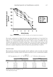
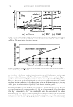
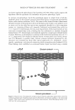
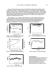
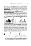
2003 ANNUAL SCIENTIFIC MEETING 225 Fig. 3: Extraction of Langerhans 's cell and melanocyte from epidennis ground. View on dermal components Glycosaminoglycans Fibroblasts are dennal cells which synthesize fibers and ground substance. By following the glycosaminoglycans spreading in fibroblasts, we can visualize distinctly a secretion area of chondroirin sulfate around the nucleus and the pericellular area (Fig. 4). Confocal microscopy acquisition gives us serial sections for reconstructing 3D fibroblastic representation and for measuring volume and area. Transparency effect on external fibroblastic "membrane" allows us to assess its secretion under LS Active Ingredient (INCi name: Mannitol (and) Cyclodwrin (and) Yea.�, Extract (and) Disodium Succinate). 3D rendering brings a functional benefit to micro-morphology developments. Fig. 4: Secretion area of chondroitin sulfate (in green) corresponds to cytoplasmic region around the nucleus (in red) and pericellular area which is made \.isible by the use of transparency effect. Elastic fibers The elastic fibers of the connective tissue form a network responsible for rhe elastic properties of skin. The organization of its network is not easily perceived with 2D images (3, 4]. The use of JD representation (Fig. 5), by a volumic rendering, allows to visualize the network organization, and the connection existing between elastic fibers. This volumic rendering corresponds to full elements, in opposition to surfacic rendering that shows only the external surface of those elements. JD visualization brings a superior dimension compared to the used traditional methods ( INCi Name of LS active ingredient: Pisum Sativum ( Pea) Extract). Fig. 5: Elastic fibers representation and its organization. In conclusion, volumic rendering offers a new approach of cutaneous structures. Extraction, segmentation, thresholding and measurements of these structures bridge the gap between histology and JD computeril.Cd representation. Then, 3D visualization allows to take into account the whole structure of skin which organizes the biological activities of cells and therefore a better evaluation of the activities from cosmetic active ingredient. By this way, 3D cosmetic universe delivers answers to many cutaneous challenges. (1] Vazquez-Duchfne MD, GiUon V, Contet-Audonneau JL, Freis 0, Perie G, Jeanmaire C, Gauch� D and Pauly G, Skin Research and Technology (abstract 53, ISBS-ISSI Hamburg). 9, 185. (2003). [ll Duchowski A, Medlin E, Coumia N, Murphy H, Gramopadhye A, �air S, Vorah J, and Melloy B, Behavior Research Methods, Instruments and Computers, 34, 573-591, (2002). [3) Jeanmaire C, DanOWI Land Pauly G, British Journal of Dennatology, 145, 10-18, (2001). (4) Contet-Audonneau JL, Jeanmaire C and Pauly G, British Journal of Dermatology, 140, 1038-1047, (1999).
Purchased for the exclusive use of nofirst nolast (unknown)
From: SCC Media Library & Resource Center (library.scconline.org)