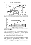
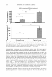
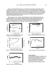
164 JOURNAL OF COSMETIC SCIENCE adsorb on the HAP surface, followed by the pellicle formation (10). The ultrastructural localization of acidic proline-rich proteins (PRPs), histatin and statherin, has been demonstrated to exist in the acquired pellicles (11). A group of PRPs is one of the major organic compounds in saliva (12). These proteins selectively adsorb on tooth surfaces (10) and delay acid diffusion (13), and, at the same time, prevent epitactic crystallization on the enamel surface (14). A review (15) has gone into detail about saliva and the dental pellicle. A number of researchers have previously studied the pellicle by a variety of analytical techniques such as infrared spectroscopy (IR) (16), X-ray photoelectron spectroscopy (XPS) (17), laser microscopy (18), auger electron spectroscopy (19), wavelength dispersive spectroscopy (20), and ellipsometry (21). Among those techniques, XPS is useful in characterizing the outermost surface of materials. The previous XPS study, however, determined only the amount of limited elements like Ca, P, 0, C, and N on the saliva-coated enamel (17). Amino acid analysis has also been performed (9,12,22), whereby the composition of a two-hour pellicle was characterized by a high content of serine, glycine, and glutamic acid instead of proline, which is a major amino acid in the whole saliva. Nevertheless, amino acid analysis has given little information about specific proteins adsorbed on the dental enamel or about the adsorption mechanism. In the present in situ study, we have confirmed that the salivary proteins selectively adsorb on HAP. However, we primarily had been interested in the mechanism whereby the salivary proteins adsorb on HAP and grow into the accumulated pellicle layers. Therefore, we have quantitatively investigated the in vitro-formed salivary film on HAP, and clarified the effects of some metal ions added to saliva on the formation of the film. Furthermore, the use of a quartz-crystal microbalance (QCM) mass-sensitive detection tool in the nanogram level has provided evidence that the electrostatic interactions between cationic and anionic proteins are involved in the alternate adsorption and the accumulation of the salivary film. We have used the QCM successfully for the first time in the field of oral science, and found it a powerful technique. MATERIALS AND METHODS IN SITU FORMATION OF SALIVARY FILM ON HAP DISKS An in situ device, comprised of a silicone rubber appliance and HAP disks simulating tooth enamel, was used for the generation of salivary film in the mouth. The HAP disks were sintered-pure hydroxyapatite (Ca/P molar ratio: 1.67 relative density: over 99% 7 mm in diameter and 4 mm in thickness), obtained from Asahi Optical Co., Ltd, Tokyo, Japan. The disks were polished using a rotary polisher to give a mirror finish. The degrees of roughness (Ra) measured by an atomic force microscope (AFM) were of the order of 30--40 nm. The devices were placed in the mouth of each volunteer to collect the salivary film material. During the collection no food or beverage was taken. After given times, the devices were removed from the mouths, and then the HAP disks were rinsed with distilled water, dried, and followed by examination using IR and XPS. CHARACTERIZATION OF IN SITU-FORMED SALIVARY FILM ON HAP DISKS Attenuated total reflection infrared spectroscopy (ATR-IR) was employed to characterize the products. All infrared spectra were obtained on a JEOL JIR2000 spectrometer ,,

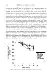
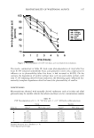
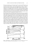
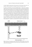
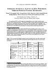
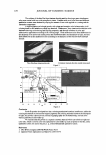
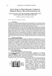
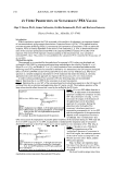
STUDY OF SALIVARY FILMS ON HYDROXYAPATITE 165 equipped with a micro-ATR attachment incorporating a sapphire prism (45°) as an internal reflection element. XPS experiments were performed with a UL V AC-PHI Model 5100 spectrometer using MgKu radiation (hv = 1253.6 eV). The utility of XPS for characterizing the surface of HAP has been demonstrated previously in our work (23,24). The Cls line at 284.6 eV was used for charge correction. SALIVA COLLECTION FOR IN VITRO STUDY Saliva samples were donated from three individuals prior to the experiments at around 9:00 a.m., before taking food or beverage. Whole mixed saliva was collected under masticatory stimulation by chewing a sheet of plastic film. Each sample of pure saliva was collected separately: parotid secretion was collected by means of Curby cups (2 5) and submandibular secretion by means of a special device (26). Those saliva samples were centrifuged at 4,000 rpm and used either undiluted or diluted with distilled water to the given concentrations. QUANTITATIVE ANALYSIS OF IN VITRO-ADSORBED SALIVARY PROTEINS ON HAP POWDER HAP powder (Wako Pure Chemical Industries, Japan) (Ca/P: 1.65 specific surface area: 8 m2 g- 1 ) was used instead of a HAP disk in this experiment. The typical adsorption experiments were carried out as follows: The HAP powder was dispersed in the saliva solutions or human albumin (Sigma, USA) standard solutions at a ratio of 30 mg:10 ml. The solutions were stirred continuously with a variable-speed magnetic stirrer (bar: 10 mm length: 4 mm p: 60 rpm) for given periods at room temperature. In the case of investigating the effects of several cations on the adsorption of protein, given amounts of CaC1 2 , MgC1 2 , and NaCl were added to the saliva or protein standard solutions. After given periods, 30 ml of distilled water was added to the dispersive solution, then centrifuged at 3,000 rpm for five minutes to remove the loosely adhered saliva. The supernatant was removed, and the solid was resuspended with 30 ml of water, then centrifuged. These procedures to remove loosely adhered saliva were repeated two more times. Then, 5 ml of HCl (0.2 mol/1) was added to dissolve the whole solids, to which 4 ml of sodium phosphate buffer (0.1 mol/1, pH 8.3) was added. The solution was mixed by a vortex mixer for five seconds. The mixture was centrifuged at 3,000 rpm for five minutes, then to 2.2 ml of supernatant was added 0.5 ml of fluorescamine acetone solution (GL Sciences, Japan) (0.03 w/v%). The solution was mixed for five seconds by a vortex mixer. After two minutes the intensity of fluorescence was measured on a Hitachi 650-60 fluorescence spectrophotometer at an excitation wavelength of 390 nm and an emission wavelength of 475 nm. The method was calibrated with the standards of the human albumin. ADSORPTION OF SALIVARY PROTEINS ON A MUCIN-COATED QUARTZ-CRYSTAL MICROBALANCE (QCM) The experiments were carried out with the QCM SF-105W (Sogo Pharmaceutical, Japan). A schematic representation of the mucin-coated quartz-crystal microbalance (QCM) is shown in Figure 1. On AT-cut quartz (9-MHz), gold electrodes (16 mm2 area)
Purchased for the exclusive use of nofirst nolast (unknown)
From: SCC Media Library & Resource Center (library.scconline.org)