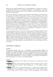
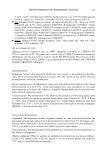
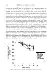
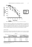
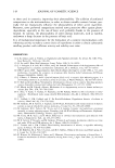
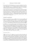
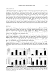
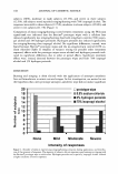
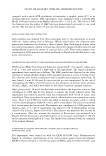
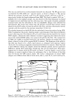
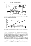
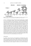
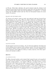
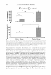
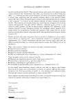
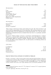
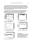
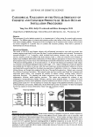
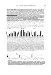
STUDY OF SALIVARY FILMS ON HYDROXYAPATITE 169 signal must be mostly responsible for the adsorption of proteins. The Cls shoulder peaks at around 287 eV arise from the carbonyl carbon. The Si2p binding energy value obtained here agrees with that of the silicates (35). QUANTITATIVE ANALYSIS OF IN VITRO-ADSORBED PROTEIN ON HAP The use of HAP disks is suitable in terms of similarity to the tooth surface, but is considered impractical for quantitative study due to the insufficient surface area for interaction. Therefore, the experiments were carried out with HAP powder for the quantitative analysis of the adsorbed proteins. The in vitro adsorption of salivary proteins on HAP powder was monitored for 120 minutes. Albumin, one of the constituents of saliva, was also used as a reference material. The time-dependent changes in the adsorp tion of both proteins are brought together for comparison in Figure 4. The initial concentration of albumin was 3,000 µg/15 ml, the saliva was diluted with the same volume of distilled water, and the amount of HAP was 30 mg. The maximum amount of adsorbed albumin (270 µg/30 mg HAP) was reached after 90 minutes, and no further increase was observed. Similarly, the adsorption plateau was observed for the saliva, although the saturation level obtained was a little lower. Next, the effect of the albumin concentration on the adsorption behavior at the collecting time of 90 minutes was investigated and is shown in Figure 5(a). The amount of adsorbed albumin reached saturation level at a concentration of around 5,000 µg/15 ml. The corresponding data for saliva are shown in Figure 5(6), where a similar pattern was obtained. The effects of Na+, Mg2+, and Ca2 + ions on the adsorption of protein were investigated, and the results at the reaction time of 90 minutes are shown in Figure 6(a) for albumin and in Figure 6(6) for saliva. Excess amounts of those cations were added to accelerate the interactions and to investigate the mechanism, though the contents of those cations in original saliva collected in the present experiments were 0.001-0.003 mol/1. The addition of each cation caused an increase in the amount of adsorbed albumin, and the Ca2 + ions showed the most significant effect. The maximum amount of adsorbed protein was reached at about 0.1 mol/1 of the cations, and then there was a gradual decrease. This was not the case, however, for the saliva in Figure 6(6). The Ca2 + ions exhibited 300 250 200 150 100 50 0L..J------------------------ 0 20 40 60 80 Reaction time(min) 100 120 Figure 4. Amount of protein adsorbed on HAP powder from the albumin solution and saliva as a function of time over periods varying from zero to 120 minutes. 0: albumin, D: saliva.

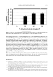
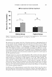
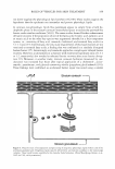
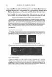
170 JOURNAL OF COSMETIC SCIENCE .s 400 -------------- J � 300� (a)� .tJ � bt)200-� � S 100- ,.C O J-4 � 0 � r:J:J '-IN 0 0 2000 1 4000 1 6000 1 8000 10000 "'d� --- Concentration of albumin( J1 g/15ml) 0.2 0.4 0.6 0.8 Dilution ratios of saliva 1.0 Figure 5. Amount of adsorbed albumin (a) and salivary proteins (b) on HAP powder in 90 minutes as functions of the concentration of albumin and the dilution ratios of saliva. The dilution ratio of unity means undiluted. characteristics similar to those in the albumin solution with regard to the increase in protein adsorption. However, the Mg2 + ions behaved oppositely to the Ca2 + ions, and Na+ ions did not considerably affect protein adsorption. The behavior in the Ca2 + and Mg2 + mixed system was closer to that in the Mg2 + system. These results reveal fundamental differences in the interaction behavior of the tested cations and suggest that the pellicle formation would be enhanced by Ca2 + ions, and that inhibited by Mg2 + ions. The agreement of the results for the three individuals has confirmed the repro ducibility of these observations, though the data are not shown here. In the present in vitro experiments, the effects of the reaction times, the protein concentrations, and the metal ions were investigated. None of the in vitro approaches, however, has resulted in a further increase in protein adsorption after reaching saturation, though the pellicle-like film actually accumulates on the enamel surface in the mouth. ADSORPTION OF SALIVARY PROTEINS ON MUCIN-COATED QCM Typical time courses of mass change (�m) on the mucin-coated QCM are shown in Figure 7. The mass increased immediately, responding to the injection of whole saliva and parotid saliva (5 ml) into distilled water (500 ml), and �m reached approximately 200 ng and 130 ng, respectively, by three-time injections for 17 minutes. On the other hand, no change for submandibular saliva occurred to any measurable degree. Then, in order to estimate the surface charge of the adsorbed salivary materials, the PEI and PSS ions were utilized as the standard cationic and anionic polyions, respectively. The
Purchased for the exclusive use of nofirst nolast (unknown)
From: SCC Media Library & Resource Center (library.scconline.org)