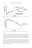
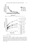
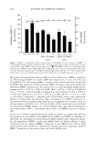
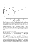
EFFICACY AND SAFETY OF DEOXY ARBUTIN 293 from light at -20°C until use. At the time of use, the compounds were further diluted in growth medium to the final concentration. For the animal study, 5 % dA, HQ and TBP were prepared in a mixture of propylene glycol, ethanol, and water at a volume ratio of 1:2:1. Solutions were kept at -20°C until use. CELL CULTURE Primary cultures of normal human melanocytes, keratinocytes, and fibroblasts were established from individual neonatal foreskins [from dark (dk) and light (lt) skin infants} that were obtained from the nursery of the University Hospital in Cincinnati after routine circumcision using a protocol approved by the University of Cincinnati Insti tutional Review Board as previously described (22). Foreskins were incubated in 0.25% trypsin for two hours at 37 ° C. The tissue was gently vortexed for 30 s to separate the dermis as a single piece and produce an epidermal cell suspension. The epidermal cells were seeded in a T-25 cm2 flask in either melanocyte or keratinocyte growth medium. Melanocytes were maintained in MCDB-15 3 growth media (Irvine Scientific, Santa Ana, CA) supplemented with 4% fetal bovine serum, 1 % antibiotic/anti-mycotic solution (Gibco, BRL, Grand Island, NY), 1 µg/ml vitamin E, 0.6 ng/ml human recombinant basic fibroblast growth factor, 5 µg/ml insulin, 0.05 µg/ml transferrin, 13 ng/ml bovine pituitary extract (Clonetics, Walkersville, MD) and 8 nM 12-O-tetradecanoylphorbol- 13-acetate. All of the above reagents were from Sigma Chemical Co. (St. Louis, MO) unless otherwise stated. The growth medium for normal human keratinocyte cultures consisted of Ml 54 basal medium (Cascades Biologicals, Portland, OR) supplemented with human keratinocyte growth supplement (Cascade Biologicals) and 1 % antibiotic/ anti-mycotic (Gibco). The dermis was vortexed and seeded into a T-25 cm2 flask with fibroblast growth medium consisting of DMEM medium (Gibco) containing 8% fetal bovine serum, 1 % glutamine (Gibco), 1 % sodium pyruvate (Gibco) and 1 % antibiotic/anti-mycotic solu tion (Gibco). All cultures were maintained in a tissue culture incubator at 3 7 ° C with 5 % CO 2 . The growth medium was routinely changed twice a week for melanocytes and fibroblasts and every other day for keratinocytes. CELL VIABILITY ASSAY Cell number (i.e., viability) was determined by direct counting. In this technique, cells were seeded at 1.3 x 10⁵ cells for melanocytes, 5 x 10⁴ cells for fibroblasts, and 5.5 x 10⁴ cells for keratinocytes, per T-12.5 cm2 flask. Cells were allowed to attach and grow for 48 hr before treatment. Cells were then treated daily with fresh growth media containing test compounds for five days. Compounds were tested at a range of concen trations in order to determine the maximum dose that did not affect the viability of human cells (experiments were performed at least in duplicate). On the sixth day, cells were detached with lX trypsin/EDTA and counted with a Coulter Counter. To deter mine the fraction of cells surviving the treatment in a specified concentration of an experimental agent, the viable cell number after each treatment was normalized using the average of the viable cell number in a control group.

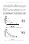
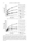
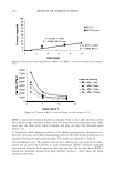
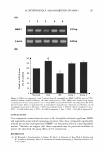
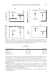
294 JOURNAL OF COSMETIC SCIENCE IN SITU (INTACT) TYROSINE HYDROXYLASE ASSAY Established melanocytes from light and dark skin were subcultivated in six-well plates at a density of 1 x 10⁵ cells/well. Cells were treated daily in triplicate for five days with fresh growth media containing different dosages of test compounds. On the fifth day cultures were assayed for tyrosine hydroxylase activity as previously described (22,23). In short, cells were fed with fresh media containing 1 µCi/ml of H 3 -tyrosine (Amersham Pharmacia Biotech, Piscataway, NJ) and the test compound. After 24-hour incubation, media from each well were collected and diluted in an equal volume of 10% (w/v) activated charcoal in a 0.1 N citric acid solution. Duplicate 1-ml aliquots of the charcoal/ media mixture were passed through a Dowex® 50Wx8-200 acidic cation 1.0-ml ex change column (Sigma-Aldrich, St. Louis, MO) followed by a 1-ml 0.1 N citric acid solution wash. The radioactivity of the tritiated water in the eluate was counted in a Packard 1900 CA liquid scintillation analyzer (Packard Instrument Company, Meriden, CT). The cultured cells in each well were harvested by trypsinization, and the cell suspension was used to determine protein and melanin content. The tyrosinase activity (DPM/24 hours/µg protein) of the cells after treatment was normalized to the activity of control cells. MELANIN AND PROTEIN CONTENT ASSAY Melanin content was determined as previously described (22). In short, cultured cells in each well were harvested with 0.2% trypsin. Cell suspensions were washed twice in phosphate-buffered saline (PBS). The cell pellets were dissolved with 100 µl Triton-X- 100 and lysates centrifuged at 13000 rpm at 4°C for 20 min. The supernatants were used to determine protein content, and pellets were used to determine melanin content. The pellets were washed with 50 µl of ethanol:ether (1:1), lysed in 100 µl of 0.2 N NaOH in 20% DMSO, and the absorbance was measured at 450 nm using a microplate reader (Bio-Rad Model 550, Japan). The melanin content of cells after treatment was expressed as µg melanin/mg protein after normalization with the control. Protein content was determined using the BCA assay (Pierce Chemical, Rockford, IL). In brief, 10 µl of supernatant was added to 200 µl of substrate (50 parts reagent A/1 part reagent B) in a 96-well plate. The plate was incubated for 30 minutes at 3 7°C and then the absorbance was measured at 570 nm in a microplate reader (Bio-Rad Model 550, Japan). The absorbance was compared with a standard curve established using known concentrations of BSA (Pierce Chemical). REVERSIBILITY ASSAY To determine if the effect of dA is reversible, inhibition of tyrosinase act1v1ty and melanin synthesis was assayed in a pulse-chase manner. Established melanocytes from dark skin donors were subcultivated in six-well plates at a density of 1 x 10⁵ cells/well. Experiments were done in triplicate using a previously determined optimal effective dosage of dA that allowed 95 % viability. After five days, a group of dA-treated and vehicle cells were assayed for the inhibition of tyrosinase activity and reduction of melanin content to demonstrate the effect of the test compound. Simultaneously, in another dA-treated group, treatment was halted and this group was fed daily with fresh
Purchased for the exclusive use of nofirst nolast (unknown)
From: SCC Media Library & Resource Center (library.scconline.org)