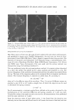
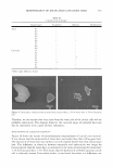
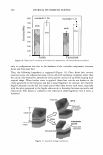
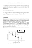
ABSTRACTS 343 determine how LA targets the follicular structure. The aim of this study was to identify the preferred route of penetration of LA and to localize the molecule, using a microautoradiographic technique associated with a compartmental approach. In an in vitro test using topical application of0.05 g of a formulation containing I% of LA, 10% of the total recovery was found in the stratum comeum and dermis after 6 h. Microautoradiographic analysis of a virtual' slide showed most of the silver grains, i.e. LA, at the hair sheath, and none in the dermal compartment, confirming the diffusion of LA through a preferred transfollicular route. These results show, for the first time on human scalp skin, that the combination of compartmental analysis and microautoradiography points to a preferred transfollicular route of diffusion of topically applied LA. Thanks to its high-resolution, microautoradiography offers the advantage of providing detailed in situ information on the delivery of LA in the skin, including the cellular location of the molecule. Charge Density Alterations in Human Hair Fibers: An Investigation Using Electrostatic Force Microscopy V. M. Longo*, V. F. Monteiro*, A. S. Pinheirot, D. Tercit, J. S. Vasconcelos*, C. A. Paskocimast, E. R. Leite*, E. Longo* and J. A Varela§ A new method for high-resolution analyses of hair surface charge density under ambient conditions is presented in this paper. Electrostatic force microscopy (EFM) is used here to analyze changes in surface charge density in virgin hair, bleached hair, and hair treated with a cationic polymer. The atomic force microscopy technique is used concomitantly to analyze morphological changes in hair roughness and thickness. . The EFM images depict exactly how the polymer is distributed on the surface of the hair fiber. The EFM's powerful analytical tools enabled us to evaluate the varying degrees of interaction between the hair fiber surface charge density and the cationic polymer. The surface charge density and the polymer's distribution in the hair fibers are presented in the light of EFM measurements. Release of Antimicrobial Actives From Microcapsules by the Action of Axillary Bacteria L. Kromidas*, E. Perriert, J. Flanagan*, R. Rivero* and I. Bonnett We describe the use of unique microcapsules that may be degraded by the action� of bacteria. These microcapsules are approximately 35 µm in diameter, are composed of natural protein, and may be filled with a variety of actives. We describe the use of antimicrobial actives such as famesol and methylparaben to demonstrate that their release by the degradative actions of axillary bacteria such as Corynebacterium minutissimum, C. urealyticum, and Staphylococcus epidermidis leads to their demise. These microcapsules may be used in consumer products such as deodorants and antiperpirants that may, under actual use conditions, control malodor.
Purchased for the exclusive use of nofirst nolast (unknown)
From: SCC Media Library & Resource Center (library.scconline.org)