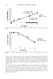
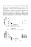
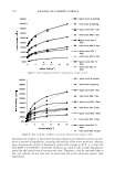
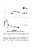
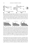
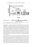
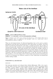
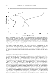
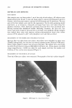
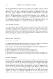
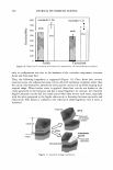
EFFICACY AND SAFETY OF DEOXYARBUTIN 295 growth media for an additional five days. At the same time, one of the vehicle-treated group had vehicle treatment halted and this group was fed daily for another five days with fresh growth media. On day 11, the four remaining groups (vehicle-treated for ten days, dA-treated for ten days, dA-treated for five days and then untreated for five days, and vehicle-treated for five days and then untreated for five days) were assessed for tyrosinase activity and melanin content. The same experimental procedure was per formed for HQ and TBP. XENOGRAFTING Xenografts were developed using a protocol approved by the Cincinnati Children's Hospital Medical Center IACUC with animal welfare assurance. Female ICR-SCID mice (Taconic, NY) kept under pathogen-free conditions (Cincinnati Children's Hospital Research Foundation, Cincinnati, OH) were shaved with an electric clipper to remove the dorsal hair. The mice were anesthetized by isofluorane/oxygen (3%/0.8 liter). The dorsal site was cut to produce a wound bed of approximately 2.0-3.0 cm in diameter. Fresh split-thickness cadaveric skin (U.S. Tissues and Cells, Cincinnati, OH) from a Caucasian donor was sutured in place with a reversed cutting precision monofilament PS-3, 6-0 (Moore Medical, CT). Grafts were left untreated for two months, during which time hyperpigmentation occurred. The degree of hyperpigmentation was assessed weekly using a microdigital image obtained from the Charm View™ (Moritex, Japan) surface optical imaging system. The treatment phase was initiated when no further increase in pigmentation was observed. Animals were balanced into three mice per group according to their L values among four treatment groups [deoxyarbutin (dA), hydro quinone (HQ), 4-tertry butyl phenol (TBP), and the vehicle-treated control group}. Treatments were topically applied at a 5 % (w/v) concentration in propylene glycol, ethanol, and water at a volume ration of 1:2:1, at 12.5 µ1/2 cm2 , five days per week, for eight weeks. The treatment sites were assessed on a biweekly basis for the degree of pigmentation using the Charm View™ system. This photographic system took enlarged digital images of the treatment sites, and then with Universal Serial Bus (USB) capture equipment, the images were transferred to a computer. Subsequently, the color param eters for these images (L, a, b) were obtained by using Adobe® Photoshop® software (Adobe Systems Inc., San Jose, Calif.). IMAGE ANALYSIS By using a histogram function in Adobe® Photoshop® software (Adobe Systems Inc.), the color or tonal range of the digital image can be evaluated in either RGB or L, a, b mode. This software allows the desired region of the image to be selected and analyzed. This includes mean, standard deviation, median, and the number of pixels of each color parameter. The L * a* b system is an international standard system, recommended by the CIE (Commission Internationale de I'Eclairage) in 1976 for skin color assessment. In this system, L* a* b color consists of a lightness component (L*), which ranges from Oto 100, and two chromatic components: a* (from green to red) and b* (from blue to yellow).

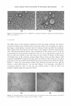
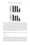
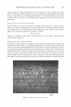
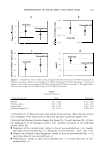
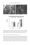
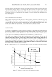
296 JOURNAL OF COSMETIC SCIENCE Both a* and b* range from -120 to + 120 (Adobe® Photoshop® 7.0 User Guide for Windows® and Macintosh). Adobe® Photoshop® uses values of 0 to 255 to characterize L, a, and b values of the selected image. L is the luminance. It gives the relative brightness from total black (L=0) to total white (L=255). The a value represents the balance between green (a=0) and red (a=255). The b value represents the balance between blue (b=0) and yellow (b=255). In this report, the L value was used to assess the lightening effect of the applied treatment and the a value was used to assess the occurrence of irritation (redness) as a result of the treatment. The L and a values obtained from Adobe® Photoshop® were converted to L * and a* values of the CIE color model by using the following formulas (24): HISTOLOGY AND IMAGE ANALYSIS L* = (L/255) (100) a*= (240a/255) - 120 (1) (2) Biopsies at the eight-week time point were processed for histology and stained with hematoxylin and eosin (H&E) and Fontana-Mason with nuclear fast red counterstain (F&M) by the Dermatopathology Laboratory in the Department of Dermatology at the University of Cincinnati. Three sections per biopsy and five different areas for each section were analyzed as follows: Images of the H&E and F&M sections were captured by a Spot Insight 4 megapixel-digital camera and Spot Imaging Software version 3.2 (Diagnostic Instruments, Inc., Burroughs, Sterling Heights, MI) attached to a light microscope using the 20 x objective. The H&E sections were analyzed for the presence of inflammation or aberrant morphology. The F&M sections were used to assess the percent of melanin per epidermal area. Fifteen images from tissues sections of each group were analyzed. For each image, the stratum corneum and the dermis were manually extracted using Adobe® Photoshop® version 7.0. Then each image in .tif format was loaded in the MATLAB® program, version 6, in RGB mode. The algorithms written in MATLAB® evaluated the red color along the (X,Y) coordinators, and saved these (X,Y) coordinators for later use in calculating epidermal size. The amount of melanin in the tissue was subsequently assessed from a duplicate image in which the original image was converted to a gray scale. Prior to a thresholding process, the red pixels that corre sponded to the saved (X,Y) coordinators were removed. This was accomplished by changing these (X,Y) coordinators to white (R, G, B = 255). The total number of black pixels corresponded to the amount of silver-stained melanin, whereas the sum of the black and red pixels represented the size of the epidermis. Finally, percent melanin in the epidermis was calculated by dividing melanin by the epidermal size and multiplying by 100. CLINICAL TRIAL A human clinical trial was performed over a six-week period with 25 male and female subjects, ages 18-60, with Fitzpatrick skin types of III or IV. Three skin sites on the back of each subject were exposed for 10-20 minutes daily for seven consecutive days to UV light from a tanning bed (Cosmolux AS bulbs emitting 2.6% UVB at 260-320 nm and 97.4% UV A at 320-400 nm). At the end of the tanning regimen, one of each of the
Purchased for the exclusive use of nofirst nolast (unknown)
From: SCC Media Library & Resource Center (library.scconline.org)