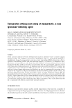
342 JOURNAL OF COSMETIC SCIENCE Ethnic Skin Types: Are There Differences in Skin Structure and Function? AV. Rawlings People of skin of colour comprise the majority of the world's population and Asian subjects comprise more than half of �he total population of the earth. Even so, t�e literature on the characteristics of the subjects with skin of colour is limited. Several groups over the past decades have attempted to decipher the underlying differences in skin structure and function in different ethnic skin types. However, most of these studies have been of small scale and in some studies interindividual differences in skin quality overwhelm any racial differences. There has been a recent call for more studies to address genetic together with phenotypic differences among different racial groups and in this respect several large-scale studies have been conducted recently. The most obvious ethnic skin difference relates to skin colour which is dominated by the presence of melanin. The photoprotection derived from this polymer influences the rate of the skin aging changes between the different racial groups. However, all racial groups are eventually subjected to the photoaging process. Generally Caucasians have an earlier onset and greater skin wrinkling and sagging signs than other skin types and in general increased pigmentary problems are seen in skin of colour although one large study reported that East Asians living in the U.S.A. had the least pigment spots. Induction of a hyperpigmentary response is thought to be through signaling by the protease-activated receptor- 2 which together with its activating protease is increased in the epidermis of subjects with skin of colour. Changes in skin biophysical properties with age demonstrate that the more darkly pigmented subjects retaining younger skin properties compared with the more lightly pigmented groups. However, despite having a more compact stratum corneum (SC) there are conflicting reports on harrier function in these subjects. Nevertheless, upon a chemical or mechanical challenge the SC harrier function is reported to be stronger in subjects with darker skin despite having the reported lowest ceramide levels. One has to remember that barrier function relates to the total architecture of the SC and not just its lipid levels. Asian skin is reported to possess a similar basal transepidermal water loss (TEWL) to Caucasian skin and similar ceramide levels hut upon mechanical challenge it has the weakest harrier function. Differences in intercellular cohesion are obviously apparent. In contrast reduced SC natural moisturizing factor levels h�ve been reported compared with Caucasi�n and African American skin. These differences will contribute to differences in desquamation hut few data are available. One recent study has shown reduced epidermal Cathepsin L2 levels in darker skin types which if also occurs in the SC could contribute to the known skin ashing problems these subjects experience. In very general terms as the desquamatory enzymes are extruded with the lamellar granules subjects with lowered SC lipid levels are expected to have lowered desquamatory enzyme levels. Increased pores size, sehum secretion and skin surface microflora occur in Negroid subjects. Equally increased mast cell granule size occurs in these subjects. The frequency of skin sensitivity is quite similar across different racial groups hut the stimuli for its induction shows subtle differences. Nevertheless, several studies indicate that Asian skin maybe more sensitive to exogenous chemicals probably due to a thinner SC and higher eccrine gland density. In conclusion, we know more of the biophysical and somatosensory characteristics of ethnic skin types hut clearly, there is still more to learn and especially about the inherent underlying biological differences in ethnic skin types. An Interlahoratory Comparison of Methods Used to Assess Antioxidant Potentials J. Buenger*, H. Ackermann•, Mehling!:, I. Pfitzner§, K.-A. Schroeder1[ and U. Wollenweber"'* A. Jentzscht, A. Reiffen*, K.-R. Many analytical methods are used to measure the antioxidative activity of substances yet little is known about the comparability of the test results between laboratories. After an initial evaluation of a broad range of methods conducted by one laboratory, the 2,2- diphenyl-1-picrylhydrazyl (DPPH) assay, the trolox equivalent antioxidant capacity (TEAC) assay, the lipid assay (or 2,2'-azohis(2-aminepropane) (ABAP) assay) and the thiobarhituric acid (TBA) assay were selected to be evaluated in the interlaboratory study. The antioxidative potentials of trolox, tocopherol, lipochroman-6, ascorbic acid, 4-methyl-hrenzcatechin, and/or 3,5-di-tert-butyl-4-hydroxytoluene (BHT) were assessed using each of the methods. These methods were then evaluated in respect of their reproducibility and classification properties. Based on the results of this study, the DPPH assay followed by the TEAC assay yielded the best results based on reproducibility and sensitivity both within one laboratory and between laboratories. The results of the interlahoratory study were then compared with the single center results obtained from the commercially available photochemolumiescence (PCL) kit. To assess the transferability of chemical data to biological systems, they were also compared with the single center results obtained using the cell-based Dichlorodihydrofluoresceine (DCFH) assay. Transfollicular Delivery of Linoleic Acid in Human Scalp Skin: Permeation Study and Microautoradiographic Analysis V. Raufast and A. Mavon There are a large number of studies on the pharmacological activity of lineolic acid (LA) on the skin however, very little work has been carried out to

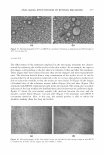
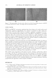
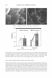
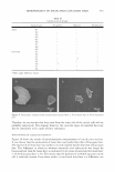
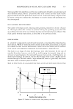
ABSTRACTS 343 determine how LA targets the follicular structure. The aim of this study was to identify the preferred route of penetration of LA and to localize the molecule, using a microautoradiographic technique associated with a compartmental approach. In an in vitro test using topical application of0.05 g of a formulation containing I% of LA, 10% of the total recovery was found in the stratum comeum and dermis after 6 h. Microautoradiographic analysis of a virtual' slide showed most of the silver grains, i.e. LA, at the hair sheath, and none in the dermal compartment, confirming the diffusion of LA through a preferred transfollicular route. These results show, for the first time on human scalp skin, that the combination of compartmental analysis and microautoradiography points to a preferred transfollicular route of diffusion of topically applied LA. Thanks to its high-resolution, microautoradiography offers the advantage of providing detailed in situ information on the delivery of LA in the skin, including the cellular location of the molecule. Charge Density Alterations in Human Hair Fibers: An Investigation Using Electrostatic Force Microscopy V. M. Longo*, V. F. Monteiro*, A. S. Pinheirot, D. Tercit, J. S. Vasconcelos*, C. A. Paskocimast, E. R. Leite*, E. Longo* and J. A Varela§ A new method for high-resolution analyses of hair surface charge density under ambient conditions is presented in this paper. Electrostatic force microscopy (EFM) is used here to analyze changes in surface charge density in virgin hair, bleached hair, and hair treated with a cationic polymer. The atomic force microscopy technique is used concomitantly to analyze morphological changes in hair roughness and thickness. . The EFM images depict exactly how the polymer is distributed on the surface of the hair fiber. The EFM's powerful analytical tools enabled us to evaluate the varying degrees of interaction between the hair fiber surface charge density and the cationic polymer. The surface charge density and the polymer's distribution in the hair fibers are presented in the light of EFM measurements. Release of Antimicrobial Actives From Microcapsules by the Action of Axillary Bacteria L. Kromidas*, E. Perriert, J. Flanagan*, R. Rivero* and I. Bonnett We describe the use of unique microcapsules that may be degraded by the action� of bacteria. These microcapsules are approximately 35 µm in diameter, are composed of natural protein, and may be filled with a variety of actives. We describe the use of antimicrobial actives such as famesol and methylparaben to demonstrate that their release by the degradative actions of axillary bacteria such as Corynebacterium minutissimum, C. urealyticum, and Staphylococcus epidermidis leads to their demise. These microcapsules may be used in consumer products such as deodorants and antiperpirants that may, under actual use conditions, control malodor.
Purchased for the exclusive use of nofirst nolast (unknown)
From: SCC Media Library & Resource Center (library.scconline.org)