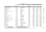
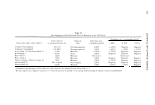
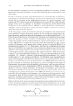
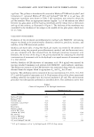
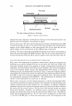
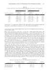
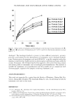
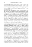
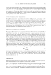
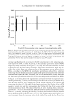
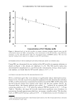
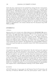
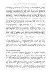
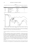
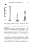
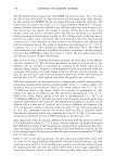
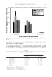
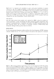
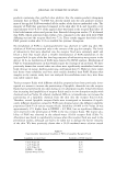
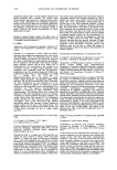
TRANEXAMIC ACID IN HYDROGEL PATCH FORMULATIONS 219 a gel base. The gel base in formulation B consisted of Methocel® E4M and Acrylax®, and formulation C contained Methocel® E50 and Carbopol® 980 NF in its gel base. All important ingredients were shown in Table I. All ingredients were mixed to obtain the gel-like mixture. Then, an appropriate amount (approx. 5 g.) of the mixture was added between two glass plates with the backing membrane and the release liner at the bottom and top of the contents, as illustrated in Figure 3. The thickness of the membrane was controlled by the thickness of two ridges in the middle of two glass plates, which were set at 2 mm. VALIDATION OF THE METHOD Evaluation of the developed spectrofluorimetric method with NDA/CN- derivatizing reagent was based on the proportionality (linearity), selectivity, precision, accuracy, and stability of the CBI-derivatized product. Specificity and matrix effect. A drug-free blank gel matrix was tested for the presence of interference using the proposed spectrofluorimetric method, and the fluorescent inten sity was compared with that obtained from the hydrogel-containing tranexamic acid. Similarly, the recovery of the drug in the gel was examined by comparing the observed concentrations of tranexamic acid in the hydrogel to an equal amount of tranexamic acid in a neat solution. Stability. Stability of CBI-derivative of tranexamic acid (50.4 µg/ml) was assessed by reacting standard tranexamic acid solution with NDA/CN- under ambient conditions. The fluorescent intensity of the CBI-derivative was monitored along with the intensity obtained form the reagent blank at appropriate Aex and Aem for 30 min. Linearity. The calibration curves consisted of the six concentrations: 8.4, 16.8, 33.6, 50.4 67 .2, and 84.0 µg/ml of tranexamic acid. A 50-µl amount of one of the above-mentioned working solutions was spiked into the reaction medium (1.85 ml). The derivatization procedure was subsequently performed as described above. The calibration curves were Table I Chemical Components of the Hydrogel Formulations Amount (g in 5 g) Component A B C Methocel ® E4M 0.75 0.70 Methocel ® E 5 0 0.59 Distilled water 3.10 2.63 2.16 Tranexamic acid powder* 0.21 0.21 0.21 Cosolvent 0.25 0.21 0.21 Preservative 0.01 0.02 0.02 Phenoxy ethanol 0.04 0.04 0.04 8%w/w DC7-9245 0.17 0.17 50%w/w DC7-9245 0.34 DC 200/350 0.17 0.17 10%w/w PVP K90 0.47 0.17 30%w/w Acrylax® 0.68 6% w/w Carbopol® 980 NF 1.26 * 0.21 g of tranexamic acid in 5 g hydrogel equals 4.20% of drug content.

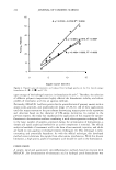
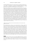
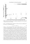
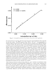
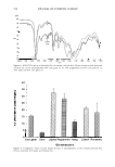
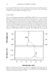
220 JOURNAL OF COSMETIC SCIENCE Glass plate Backing membrane Hydrogel Glass plate The ridge setting up thickness of hydrogel Figure 3. Diagram of patch making. obtained by linear regression the fluorescent intensity of the derivatized product was plotted versus the tranexamic concentration in µg/ml. Precision and accuracy. The intra- and interday precision (relative standard deviation, RSD % ) and the interday accuracy (% recovery) of the assay procedure were determined by analysis of five spiked samples at each concentration level on the same day and one spiked sample at each concentration on five different days, respectively. Sensitivity. The limit of quantification (LOQ) was defined as the lowest concentration at which the precision expressed by RSD was lower than 2%, the accuracy expressed by % recovery was between 98% and 102%, and the signal-to-noise ratio was better than 10. ANALYSIS OF TRANEXAMIC ACID IN HYDROGEL PATCH FORMULATION Drug content. The examination of tranexamic content from a patch was carried out by dissolving an accurately weighed (0.20 g) cut gel patch into 100.0 ml of PBS. The tranexamic hydrogel patches were prepared to contain a homogeneous distribution of tranexamic acid in the amount of 0.042 gig of gel. Therefore, the content of tranexamic acid in the dissolved sample solution was approximately 0.084 mg/ml. In order to solubilize the gel, the mixture had to be kept at least 12 hours for complete solubili zation before the volume of the solvent was adjusted. Every sample was diluted 50 times with PBS in order to fit within the range of the calibration curve. The diluted sample was subsequently derivatized with NDA/CN-. The determination of tranexamic acid content in each formulation was carried out in triplicate. Release study of tranexamic acid from hydrogel preparations. A side-by-side horizontal diffu sion cell was utilized for the release experiment. Two tranexamic acid patch samples were carefully mounted back-to-back between the two chambers. A hydrated cellophane membrane was placed on top of each hydrogel patch, and served as the barrier between the matrix and the receiver medium. The top side of the hydrogel patches was placed facing both chambers. Therefore, two diffusion systems consisting of two hydrogel patches were obtained by using one side-by-side horizontal diffusion unit. The diffusion surface area of a horizontal cell was 1.54 cm2 . The receiver compartment was filled with 2.5 ml of 0.15 M PBS, pH 7.4, which was continuously stirred. The temperature of the
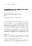
Purchased for the exclusive use of nofirst nolast (unknown)
From: SCC Media Library & Resource Center (library.scconline.org)