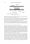
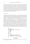
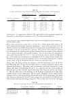
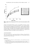
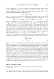
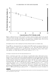
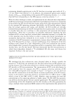
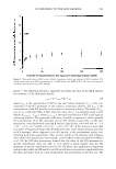
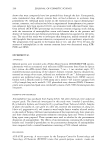
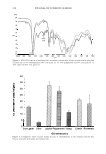
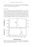
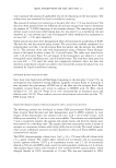
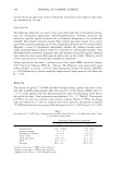
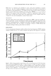
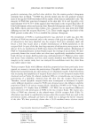
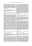
240 JOURNAL OF COSMETIC SCIENCE penetrating through aqueous pores in the SC that have an average pore radius of 3 3 ± 5 A (see Table I and reference 13). In addition, the normalized aqueous pore number density, (sl'T) 00rmal , corresponding to the aqueous SCI contacting solution, 2 ± 1, is less than half that corresponding to the SDS aqueous contacting solution, 7 ± 1. With the above findings in mind, our explanation for the observed dose independence of SCI skin penetration and associated skin barrier perturbation is based on a comparison of the radius of an SCI micelle with the average radius of the skin aqueous pores induced by the aqueous SCI contacting solutions. Specifically, the SCI micelle radius is larger than the average aqueous pore radius, and therefore, the SCI micelles are sterically hindered from penetrating into the SC through these skin aqueous pores. As a result, SCI in micellar form does not contribute to skin penetration and associated skin barrier perturbation. 3 Note that in providing our plausible explanation regarding the skin mildness of SCI, we have implicitly assumed that a surfactant monomer, or a micelle, has to first penetrate into the skin barrier in order to induce skin barrier perturbation, an assumption that has been validated by in vivo studies (1-10,14-16). Because the con centration of the SCI monomers is approximately constant above the CMC (1 mM) while that of the SCI micelles increases, a natural manifestation of our explanation of the skin mildness of SCI should be a dose independence of SCI skin penetration (see below), which should reflect primarily the penetration of SCI in monomeric form, rather than in micellar form, into the skin barrier. We have determined that SCI skin penetration is indeed dose-independent through a 14 C-radiolabeled SCI skin assay, and discuss this important finding in the next section. VERIFICATION OF OUR EXPLANATION FOR THE SKIN MILDNESS OF SCI USING THE SCI SKIN RADIOACTIVITY ASSAY We developed the skin radioactivity assay discussed above to directly quantify the amount of 14 C-SCI that can penetrate into the skin barrier from an SCI aqueous con tacting solution. Use of this assay allowed us to directly measure the contribution of the SCI micelles to SCI skin penetration. The concentrations of SCI in the skin barrier (in wt%) resulting from the exposure of p-FTS to aqueous contacting solutions of SCI (0.2-200 mM) correspond to the triangles in Figure 5. In Figure 5, one can clearly see that the SCI concentration in the skin barrier increases significantly as the SCI concen tration in the aqueous contacting solution is increased from 0.2 mM to about 5 mM, which is close to the CMC of SCI (1 mM). However, upon increasing the total SCI concentration in the contacting solution to higher values, 50-200 mM, the concentra tion of SCI in the skin barrier does not increase significant! y. We quantified the relative contributions of the SCI monomers and the SCI micelles to SCI skin penetration by using a multiple linear regression (MLR) analysis of the experimental data presented in 5 The CMC of SCI is 1 mM, which is much lower than the CMC of SDS (8.7 mM), which is a well-known harsh skin agent. Therefore, at a surfactant concentration above the CMC, SCI has a much smaller concen tration of monomers that come in contact with the skin than SDS. Hence, not only does SCI form larger micelles (relative to SDS) that cannot penetrate into the SC, but it also has a lower concentration of monomers contacting the skin (relative to SDS) that can penetrate into the SC and induce skin barrier perturbation.

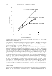
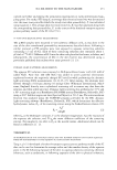
SCI MILDNESS TO THE SKIN BARRIER 241 3 2.5 � � ·c � 2 h CMC rLJ. � 1.5 f-1 .s -� � 1 u = I � I = nfJ u 0.5 I I I I I u rLJ. 0 J I 0 50 100 150 200 Total SCI Concentration in the Aqueous Contacting Solution (mM) Figure 5. Skin penetration of SCI in vitro induced by aqueous contacting solutions of SCI (triangles). The dotted vertical line at an SCI concentration of 1.0 mM denotes the CMC of SCI. The error bars represent standard errors based on six p-FTS samples. Figure 5. The following relation is applicable and forms the basis of the MLR analysis (see reference 11 for additional details): (6) where Cskin is the concentration of SCI in the skin barrier (mmol/g), Cmon is the con centration of the SCI monomers in the aqueous contacting solution, and Cmic is the concentration of the SCI micelles in the aqueous contacting solution. The value of Cmon was set to 1 mM (the CMC of SCI) while the value of Cmic was obtained using a mass balance (CscI,total - cman J where CsCI,total is the total concentration of SCI in the aqueous contacting solution. The regression coefficients, a and 13, in equation 6, which quantify the contributions of an SCI monomer and an SCI micelle, respectively, to SCI skin penetration, were determined using MLR analysis. Specifically, we found that: a = 1.6 x 10- 2 ± 8.4 x 10⁻⁴ and 13 = 6.8 x 10- 5 ± 9.8 x 10- 6 (R2 =0.93). Since a is more than two orders of magnitude larger than 13 (a/13=238), this result clearly demonstrates that an SCI monomer, when compared to an SCI micelle, is the predominant species con tributing to SCI skin penetration. This, in turn, results in the observed dose indepen dence of SCI skin penetration. Interestingly, a similar MLR analysis performed by Moore et al. (11) for SDS skin penetration showed that the value of the SDS monomer-to micelle contribution ratio, our a/l3, is 3.25, which is much smaller than the SCI monomer-to-micelle contribution ratio of 238. Therefore, this quantitative comparison indicates that unlike an SDS micelle, an SCI micelle does not penetrate significantly into the skin, and hence leads to a dose-independent skin penetration in the SCI case. The
Purchased for the exclusive use of nofirst nolast (unknown)
From: SCC Media Library & Resource Center (library.scconline.org)