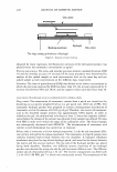
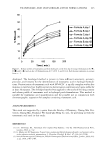
232 JOURNAL OF COSMETIC SCIENCE p-FTS sample was mounted in the diffusion cell with the SC facing the donor com partment. Both the donor and the receiver compartments were filled with PBS, and the p-FTS sample was left to hydrate for one hour before the beginning of the experiment to allow the skin's initial barrier property to reach steady state. At this point, the skin electrical current across the p-FTS sample was measured, and only p-FTS samples with an initial skin current 3 µA were used in the permeation studies (11,12,28,31). The PBS in the donor compartment was then replaced with 1.5 ml of an SCI aqueous solution. The diffusion cell was then transferred to a temperature-controlled oven, with the temperature set at 3 5 °C to prevent SCI precipitation from the contacting solution in the donor compartment of the diffusion cell (42). The donor compartment of the diffusion cell was covered by para-film to prevent water evaporation at this temperature. The SCI aqueous contacting solution in the donor compartment contacted the p-FTS sample for five hours (11,12). Subsequently, the contacting solution was removed and the donor compartment and the p-FTS sample were rinsed four times with 2 ml of PBS to remove any trace chemical left on the skin surface and in the donor compartment. The receiver compartment was stirred with a magnetic stirrer at a speed of 400 rpm through out the experiment to eliminate permeant bulk concentration gradients. Following the SCI aqueous contacting solution treatments of the skin, the p-FTS samples in the diffusion cells were exposed to a contacting solution of 3 H-radiolabeled mannitol in PBS (1-10 µCi/ml) for 24 hours (24,27,31). Throughout these experiments, solution samples were withdrawn from both the receiver (r) and the donor (d) compart ments every two hours, and the concentrations of the permeant (mannitol) in the two compartments (C r and C d) respectively) were measured using a liquid scintillation counter (Packard, Sheldon, CT). When the transport of mannitol attained steady state, the mannitol skin permeability, P) was calculated as follows (24,27 ,31): p = __( l d(CrVr) ) (1) ACd dt where Vr is the volume of the receiver compartment, A = (1.77 cm2) is the area of the SC exposed to the mannitol solution in the donor compartment, and t is the exposure time. IN VITRO SKIN ELECTRICAL CURRENT AND SKIN ELECTRICAL RESISTIVITY MEASUREMENTS During each skin permeation experiment, two Ag/AgCl electrodes (E242, In Vivo Metrics, Healdsburg, CA) were placed in the donor and receiver compartments to measure the electrical current and the electrical resistivity across the p-FTS sample (31). A 100 m V AC voltage (RMS) at 10 Hz was generated by a signal generator (Hewlett Packard, Atlanta, GA), and was applied across the two electrodes for 5 s. The electrical current across the skin was measured using an ammeter (Hewlett-Packard). This am meter was used to measure low AC currents and was accurate in the 0.1 µA range. The electrical resistance of the p-FTS sample was then calculated from Ohm's law (31). Because the measured skin electrical resistance is the sum of the actual skin electrical resistance and the background PBS electrical resistance, the latter was subtracted from the measured skin electrical resistance to obtain the actual skin electrical resistance. The skin electrical resistivity was then obtained by multiplying the actual skin electrical resistance by the skin area (A = 1.77 cm2). Additional details about this procedure are provided in reference 13. Skin electrical current and resistivity measurements were

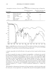
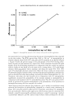
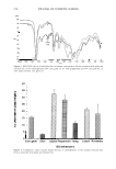
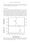
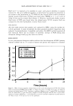
SCI MILDNESS TO THE SKIN BARRIER 233 carried out before and during the permeation experiments at each predetermined sam pling point. For each p-FTS sample, an average skin electrical resistivity was determined over the same time period for which the steady-state skin permeability, P, was calculated using equation 1. This average skin electrical resistivity, R, was then analyzed along with the corresponding skin permeability, P, in the context of the hindered-transport aqueous porous pathway model of the SC (26,27 ,31). IN VITRO SKIN RADIOACTIVITY MEASUREMENTS The p-FTS samples were mounted in vertical Franz diffusion cells, as was done in the case of the skin transdermal permeability measurements described above. Following a similar protocol, p-FTS samples were now exposed to aqueous contacting solutions containing 1. 5 ml of SCI. Each of these contacting solutions also contained about 1 µCi/ml of 14 C-SCI. Diffusion of SCI into the skin took place for five hours, as before, and subsequently, the concentration of SCI in the skin barrier was determined using a previously published skin radioactivity assay protocol (11-13 ). DYNAMIC LIGHT-SCATTERING MEASUREMENTS The aqueous SCI solutions were prepared in Millipore-filtered water with 100 mM of added NaCl. Note that 100 mM NaCl was added to screen potential electrostatic repulsions between the negatively charged SCI micelles while performing the dynamic light-scattering (DLS) measurements (11,13,35-37). After mixing, the solutions were filtered through a 0.02-µm Anotop 10 syringe filter (Whatman International, Maid stone, England) directly into a cylindrical scattering cell to remove any dust from the solution, and then sealed until use. Dynamic light scattering was performed at 3 5 ° C and a 90° scattering angle on a Brookhaven Bl-200SM system (Brookhaven, Holtsville, NY) using a 2017 Stabilite argon-ion laser (Spectra Physics) at 513.5 nm. The autocorrelation function was analyzed using the CONTIN program provided by the BIC dynamic light-scattering softw�re (Brookhaven, Holtsville, NY), which determines the effective hydrodynamic radius, Rh, of the scattering entities using the Stokes-Einstein relation (35): - kBT Rh= -------== (2) 6TI11D where k B is the Boltzmann constant, Tis the absolute temperature, 11 is the viscosity of the aqueous salt solution, and D is the mean diffusion coefficient of the scattering entities. For simplicity, we refer to R h as the micelle radius. Additional details can be found in references 11-13. THEORETICAL DETERMINATION OF THE AVERAGE RADIUS AND THE NUMBER DENSITY OF THE SKIN AQUEOUS PORES IN SKIN EXPOSED TO THE AQUEOUS SCI CONTACTING SOLUTIONS Tang et al. (31) developed a hindered-transport aqueous porous pathway model of the SC that can be used to determine the average radius and the number density of the aqueous pores in the SC following exposure of the skin to aqueous solutions containing chemical enhancers, such as surfactants, or following exposure of the skin to physical enhancers,
Purchased for the exclusive use of nofirst nolast (unknown)
From: SCC Media Library & Resource Center (library.scconline.org)