258 JOURNAL OF COSMETIC SCIENCE The full-thickness frozen human skin from NDRI was stored at about -80 ± 2°C until use. On the day of the study, the skin was thawed at room temperature. After thawing, the skin was placed in HHBSS. The fat was trimmed from the underside of the skin. The human skin was washed once with a 1 % v/v liquid detergent (Palmolive® "Original" formula) solution, rinsed with distilled water, and then patted dry with a paper towel. The porcine skin was shaved with clippers to remove all hair from the surface, then washed, rinsed, and dried as described above. The skin was positioned on a styrofoam block and secured with hypodermic needles. A 180-3 30 µm section of the skin was cut from the top surface using a dermatome. Discs of dermatomed skin were obtained using a steel punch (17-mm diameter) and then each disc was mounted in the diffusion cell with the epidermal side up. Skin surface temperature was maintained at 32° ± 2°C by circulating 36° ± 1 ° C water through the diffusion cell-holding block. The diffusion cell-mounted skin was allowed to equilibrate for at least 30 minutes, with the pumping of receptor fluid (HHBSS) at a flow rate of about 1.5 ml/h. The skin surface temperature was measured with an infrared thermometer. After at least 30 min, a 3 H-water skin-barrier integrity test of the skin in the diffusion cells was conducted (5). The 3 H-water skin-barrier integrity test of the skin in the diffusion cell was necessary to determine the integrity of the barrier (skin may be damaged during surgery, transport, or skin preparation) prior to application of the test compound. If the barrier test results indicated that the skin disc was too permeable, the skin disc was not used in the study (the historical upper limit of 3 H-water permeation through skin is 0.35% of the applied dose under the specified test conditions). PAN skin absorption was determined from a commercially available suntan product (oleaginous ointment-based product) containing D&C red no. 1 7 ("dosing vehicle" the estimated level of non-radioactive PAN in the suntan product was 1.2 x 10- 5 %). 14 C-PAN was added to the dosing vehicle in an amount to approximate 0.5 µCi radioactivity applied per diffusion cell. This represented approximately 1.8% of 14 C PAN in the dosing vehicle applied to the skin or approximately 1.2 x 10 5 g of 14C-PAN per diffusion cell. The skin was dosed with an amount of suntan product that simulates normal human use conditions. The formulation was applied to the skin in each diffusion cell at a dose rate of 1 mg/cm2 or 0.64 mg per diffusion cell (diameter 0.64 cm2 ) and allowed to remain on the skin surface for either 15 min (short-term exposure) or 24 h. The unabsorbed material was removed from the skin as described below. Percutaneous absorption studies were conducted for 24 and 72 h. The 72-h studies were conducted to determine the fate of PAN remaining in the skin after 24 h. After application of the test material, diffusion cell receptor fluid fractions were collected at 6-h intervals for a total of 24-72 h using a fraction collector. An aliquot of receptor fluid efRuent was removed, and the amount of radioactivity in each fraction was deter mined by liquid scintillation counting (Beckman LS 6500, Fullerton, CA). At the end of the exposure period, the skin was washed and rinsed to remove unabsorbed 14 C-PAN from the skin surface. Either 15 min or 24 h after application, the skin surface was washed three times with 0.1 ml of 1 % v/v liquid detergent (Palmolive® "Original" formula) solution, and then rinsed two times with 0.1 ml of distilled water pipetted onto the skin surface. During the washing procedure, the skin was gently rubbed with a cotton-tipped applicator to remove the detergent solution or water. The cotton tips were collected in a scintillation

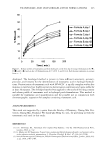
SKIN ABSORPTION OF D&C RED NO. 1 7 259 vial to measure the amount of unabsorbed test article remaining on the skin surface. The radioactivity was measured by liquid scintillation counting. The amount of radioactivity remaining in the skin after 24 or 72 h was determined. The skin discs were removed from the diffusion cell and tape stripped ten times to determine the amount of 14 C-PAN remaining in the stratum corneum. The remaining epidermal/ dermal tissue in each non-viable human skin disc was placed in a scintillation vial and dissolved in 1 ml of water and 3 ml of Scintigest® tissue solubilizer by incubation in an oven (60° ± 2°C) until dissolved. Viable porcine skin discs were homogenized when determining the metabolism of PAN. The skin disc was first minced using scissors. The pieces were carefully transferred to a polypropylene test tube, 2 ml of receptor fluid was added, and the mixture was chilled on ice. The contents of the tube were homogenized using a Polytron Tissue Homog enizer at full speed for three to four 10-15-second bursts. The tubes were rinsed twice with 1 ml of receptor fluid. A 1-ml aliquot of the homogenate was added to 3 ml of Scintigest® tissue solubilizer and 1 ml of distilled water. The vials were then placed in an oven (60° ± 2°C) until the tissue was completely dissolved. Once the skin was dissolved, scintillation cocktail was added to the vial and the amount of radioactivity was measured by liquid scintillation counting. LIPOPHILIC RECEPTOR FLUID STUDIES Since there were high levels of PAN found remaining in the skin after 24 and 7 2 h, an investigation was conducted testing different lipophilic receptor fluids to determine if they increased the partitioning of PAN out of the skin into the receptor fluid. Four lipophilic receptor fluids were tested in addition to HHBSS with 4% BSA, which included 1 %, 3%, and 6% Volpo 20 as wt% concentrations in deionized water and ethanol-water (50:50). These studies were run using human cadaver skin and the 14 C p AN dosing vehicle. HIGH-PERFORMANCE LIQUID CHROMATOGRAPHY (HPLC) ANALYTICAL METHOD The following method was developed to isolate PAN and potential PAN metabolites from receptor fluid fractions and skin homogenates prior to HPLC analysis. A 1-ml aliquot of skin homogenate was extracted one time with 3 ml of ethyl acetate, with shaking not exceeding 15 min on a wrist-action shaker. The mixture was centrifuged for 5 min to completely separate the aqueous and organic layers, and then the organic layer was removed to a centrifuge tube. The organic layer was concentrated under a stream of nitrogen to a minimum volume while being chilled in an ice bath. The small volume of ethyl acetate solubilized material was then dissolved in 200 µl of filtered HPLC grade acetonitrile for analysis. The HPLC chromatography column was a 5-µ C 18 4.6 x 250 mm (Alltech) column with a C 18 guard column. The mobile phase consisted of acetonitrile and an ammonium acetate aqueous solution (1.875 g of ammonium acetate and 2.5 ml of acetonitrile diluted to 500 ml with HPLC grade water) run under gradient conditions at 1.0 ml/min. The gradient began with a mixed eluent of 10% acetonitrile/90% aqueous phase, which changed linearly to 100% acetonitrile at 30 min. The run was continued at 100%
Purchased for the exclusive use of nofirst nolast (unknown)
From: SCC Media Library & Resource Center (library.scconline.org)