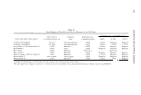
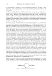
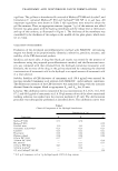
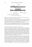
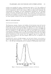
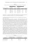
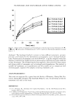
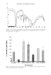
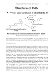
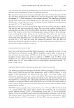
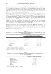
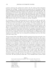
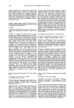
260 JOURNAL OF COSMETIC SCIENCE acetonitrile for an additional 30 min. Ultraviolet absorption of the analyte in the eluate was monitored at 256 nm. DATA ANALYSIS The following definitions are used in this study when referring to dermal/skin absorp tion and dermal/skin penetration. Skin/dermal/percutaneous absorption represents the amount of topically applied chemical that is ultimately determined to be systemically available. This would constitute receptor fluid content plus skin content if it is deter mined that material remaining in the skin ultimately partitions into the receptor fluid. Therefore, it must be determined individually whether the stratum corneum and/or viable epidermal/dermal content should be considered as systemically available. Skin/ dermal/percutaneous penetration represents the total amount of topically applied chemical that is found in the receptor fluid plus the skin at the end of a study. However, not all of this material may be systemically available for absorption. Values reported are the mean ± standard error of the mean (SEM). Statistical (Sigma Stat® Statistical Software, SPSS Inc., Chicago, IL) differences were determined using either a Student's t-test (p 0.05) or, where appropriate, analysis of variance (ANOVA, p 0.05) followed by a posteriori multiple comparisons of group means by the Tukey test (p 0.05). RESULTS The percent of applied 11C-PAN absorbed through human cadaver skin from a com mercially available suntan product after 24 h was 0.07 ± 0.004 (mean ± SEM), with 12.5 ± 2. 7 % of the applied dose that had penetrated the skin still remaining in the skin at the end of the study. Total penetration was therefore 12.6 ± 2.7 (Table I). The majority of PAN in the skin was located in the stratum corneum (Figure 2). When the dosing time was reduced to 15 min to simulate short-term exposure, the percent of applied 14 C-PAN absorbed into the receptor fluid decreased to 0.02 ± 0.002% and 11.9 ± 4.0% Table I Penetration of 11 C-PAN from a Commercially Available Suntan Product (24-h Application) after 24 and 72 h in Human Cadaver Skin Percentage of applied dose penetrated Receptor fluid Stratum corneum Epidermis and dermis Total in skin Total penetration Wash 24 h 1 0.07 ± 0.004 7.3 ± 1.4 5.2±1.4 12.5 ± 2.7 12.6 ± 2.7 95.7 ± 10.3 Recovery 108.3 ± 9.9 1 Values are the mean ± SEM of seven replicates. 2 Values are the mean ± SEM of eight replicates. * Different from 24 h, p 0.001 (Student's t-test). 72 h 2 0.17 ± 0.01* 9.7 ± 1.9 5.6 ± 0.9 15.3 ± 2.6 15.5 ± 2.6 91.5 ± 6.7 107.0 ± 6.4

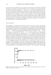
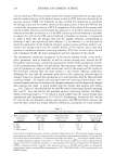
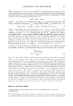
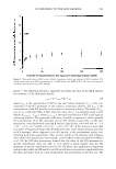
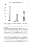
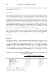
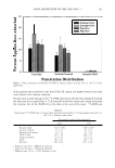
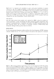
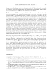
SKIN ABSORPTION OF D&C RED NO. 1 7 261 � 25 ------------------------------- ,.Q � 0 r:l'l ,.Q � r:l'l 20 0 15 � Total Skin Stratum Corneum - Cadaver-24 hr � Cavader-72 hr l\\'1111 Pig-24hr � Pig-72 hr Receptor Fluid Penetration Distribution Figure 2. Skin penetration distribution of PAN in human cadaver and pig skin 24 and 72 h after application. of the applied dose remained in the skin (Table II). Again, the highest levels in the skin were found in the stratum corneum. Because such a small amount of the 14 C-PAN penetrating the skin was absorbed through the skin into the receptor fluid, a 72-h extended study was conducted to help determine the systemic fate of the PAN left in the skin at the end of the study. 14 C-PAN was Table II Penetration of 14 C-PAN from a Commercially Available Suntan Product (15-min Application) after 24 and 72 h in Human Cadaver Skin Receptor fluid Stratum corneum Epidermis and dermis Total in skin Total penetration Wash Recovery 1 Values are the mean ± SEM of six replicates. 2 Values are the mean ± SEM of six replicates. Percentage of applied dose penetrated 24 h 1 0.02 ± 0.002 7.3 ± 2.7 4.6 ± 1.4 11.9 ± 4.0 11.9 ± 4.0 78.7 ± 8.7 90.6 ± 8.8 72 h2 0.08 ± 0.02* 9.7 ± 3.7 4.3 ± 0.9 14.0 ± 4.5 14.0 ± 4.5 75.6 ± 7.6 89.7 ± 6.8 * Different from 24 h, p 0.001 (Student's t-test).
Purchased for the exclusive use of nofirst nolast (unknown)
From: SCC Media Library & Resource Center (library.scconline.org)