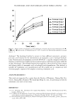
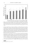
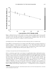
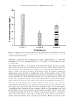
230 JOURNAL OF COSMETIC SCIENCE skin (1-23,39,41). Several factors have been proposed to explain the mildness of SCI relative to the harshness of other skin agents used in skin cleansers, including: (i) a lower critical micelle concentration (CMC) value for SCI, resulting in a lower SCI monomer activity with the skin barrier (1-3,15), (ii) reduced penetration of SCI into the skin barrier (1-5), and (iii) decreased binding of SCI to proteins and lipids in the stratum corneum (SC), which is the primary constituent of the skin barrier, resulting in a lower skin irritation response (3-9). The interactions of SCI with the SC proteins have been studied extensively (1,3,15,42). These studies have shown that one of the factors responsible for the skin mildness/ harshness for a surfactant solution is the charge density of the surfactant polar head group. For example, it has been shown that SCI has a lower charge density than SDS, and therefore binds less strongly to the SC proteins (1,42). The CMC of the SCI aqueous solution has been considered as another important factor. Specifically, Ananthapadma nabhan et al. (1) observed that SCI has a lower CMC than SDS and binds to proteins in the SC only about one-fifth as much as SDS under similar solution conditions and exposure times. The interaction of SCI with SC lipids has also been investigated to shed light on the mechanisms of SCI-induced skin barrier perturbation. Specifically, nonionic surfactants such as alkyl polyglucosides, which do not interact strongly with SC proteins, have been shown to dissolve stearic acid and cholesterol to a much greater extent than mild anionic surfactants like SCI (42). Because SCI does not selectively remove fatty acids and cholesterol from the lipid bilayers in the SC, it does not induce significant biological damage through the modification of lipid biosynthetic functions due to changes in the relative levels of various lipids in the SC (17-20). It is well accepted that surfactants have to first penetrate into the skin barrier before they can reduce the skin barrier properties. Therefore, if a formulator can minimize surfactant skin penetration, this should also minimize the ability of the surfactant to reduce the skin barrier properties. Our previous research has investigated the process of SDS skin penetration from an aqueous contacting solution by itself (11), as well as when mixed with (i) a polymer-polyethylene oxide (PEO) (11), (ii) a nonionic surfactant-dodecyl hexa(ethylene oxide) C 12 E6 (12), and (iii) a humectant-glycerol (13). It is well-known that the SDS monomers self-assemble in aqueous solution to form micelles at SDS concentrations that are greater than the CMC of SDS. Due to the hydrophilic nature of the resulting SDS micelles, they are expected to penetrate into the SC through aqueous pores that exist in the SC (11, 13 ). These aqueous pores in the SC are located in the lacunae and other aqueous regions surrounded by polar lipids in the lipoidal mortar between the corneocyte bricks that comprise the brick-and-mortar structure of the SC (24,25). We have recently shown that the SDS micelles are smaller in radius than the average radii of these aqueous pores, and therefore contribute to SDS skin penetration and induce skin barrier perturbation (11-13 ). Strong evidence that the SDS micelles do indeed contribute to SDS skin penetration is also provided by the observed dose depen dence of SDS skin penetration and barrier perturbation, which we found to increase as the total SDS concentration was increased beyond the CMC of SDS (8-13 ). In cases (i) and (ii) above, where PEO and C 12 E 6 were added separately to aqueous SDS contacting solutions, the SDS micelle radius increased relative to the aqueous pore radius, such that the larger SDS micelles became sterically hindered from penetrating into the SC through the aqueous pores (11,12). On the other hand, in case (iii), when glycerol was added to an aqueous SDS contacting solution, the average aqueous pore radius decreased relative

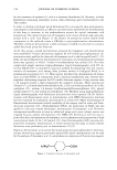
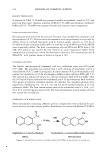
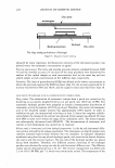
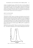
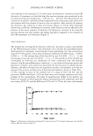
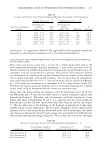
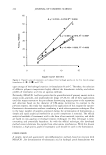
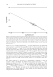
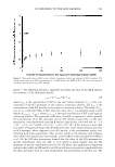
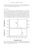
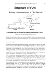
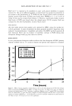
SCI MILDNESS TO THE SKIN BARRIER 231 to the SDS micelle radius, such that the SDS micelles became sterically hindered from penetrating into the SC through the smaller aqueous pores (13 ). In this paper, we hypothesize that the well-documented skin mildness of SCI is due to the inability of the SCI micelles to contribute to skin penetration and induce skin barrier perturbation. To test this hypothesis, we have determined the average radius and the number density of the aqueous pores in the SC when an aqueous solution of SCI contacts the skin. To this end, we have conducted in vitro mannitol skin permeability and skin electrical current measurements in the context of a hindered-transport aqueous porous pathway model of the SC. We have also carried out dynamic light-scattering (DLS) measurements to determine the radius of the SCI micelles present in the aqueous solution contacting the skin. Such a combined in vitro investigation can explain the well-documented in vivo and in vitro skin mildness of SCI (1-3,42) by showing that the radius of an SCI micelle is significantly larger than that of the skin aqueous pores. As a result, SCI in micellar form is unable to penetrate into the SC through these aqueous pores. Finally, we have also measured the penetration of SCI into the skin using in vitro 14 C-radiolabeled SCI skin radioactivity assays. Because only the SCI monomers can contribute to SCI skin penetration, due to the inability of the SCI micelles to do so based on the size limitation discussed above, we will show that the SCI skin penetration is dose-independent, an important finding that further validates our hypothesis. EXPERIMENT AL MATERIALS Sodium cocoyl isethionate (SCI) from BASF was provided to us by Unilever (Edgewater, NJ). 14 C-radiolabeled SCI and 3 H-radiolabeled mannitol were purchased from American Radiolabeled Chemicals (St. Louis, MO). Sodium chloride (NaCl) was purchased from Sigma Chemicals (St. Louis, MO). All these chemicals were used as received. Water was filtered using a Millipore Academic water filter (Bedford, MA). Phosphate-buffered saline (PBS) was prepared using PBS tablets from Sigma Chemicals (St. Louis, MO) and Millipore-filtered water, such that a phosphate concentration of 0.01 M and a NaCl concentration of 0.137 M were obtained at a pH of 7.2. PREPARATION OF THE SKIN SAMPLES Female Yorkshire pigs (40-45 kg) were purchased from local farms, and the skin (back) was harvested within one hour after sacrificing the animal. The subcutaneous fat was trimmed off using a razor blade, and the full-thickness pig skin was cut into small pieces (2 cm x 2 cm) and stored in a -80°C freezer for up to two months. The surfactant penetration experiments were conducted using pig full-thickness skin, referred to here after as p-FTS. IN VITRO TRANSDERMAL PERMEABILITY MEASUREMENTS Vertical Franz diffusion cells (Permegear Inc., Riegelsville, PA) were utilized in the in vitro transdermal permeability measurements (11,12,31). Prior to each experiment, a
Purchased for the exclusive use of nofirst nolast (unknown)
From: SCC Media Library & Resource Center (library.scconline.org)