422 JOURNAL OF COSMETIC SCIENCE 96-well plates for 10 min at room temperature. The absorbance at 570 nm was measured by spectrophotometry. RNA ISOLATION AND RT-PCR OF iNOS Raw 264. 7 cells were seeded at a density of 2 x 10⁶ cells into a 100-mm dish and cultured at 37°C in 5% CO2 . After one day, fresh medium containing 10% serum was added to the cells, which were then treated with LPS (lipopolysaccharide, 1 µg/ml) and the ethanol extract of Crinum asiaticum for 8 h. Total RNA was isolated from cells with TRizol (Invitrogen) according to the instructions of the manufacturer. First-strand cDNA synthesis was performed using random hexamers. The sequences of primers are as follows: 5 '-CAGTTCTGCGCCTTTGCTCAT-3' (sense) and 5 '-GGTGGTGCGG CTGGACTTT-3' (antisense) for iNOS, and 5 '-GACGTGCCGCCTGGAGAAA-3' (sense) and 5'-GGGGGCCGAGTTGGGATGG-3' (antisense) for GAPDH. The RT PCR reaction of iNOS was reverse-transcripted at 50°C for 30 min and denatured at 96 ° C for 3 min, followed by 32 cycles at 94°C for 30 sec, 60 ° c for 30 sec, and 72°C for 1 min, and then followed by an extension step cycle at 72°C for 10 min. The RT-PCR reaction of GAPDH (glyceraldehyde-3-phosphate dehydrogenase) was reverse transcripted at 50°C for 30 min and denatured at 96°C for 3 min, followed by 32 cycles at 94°C for 30 sec, 60°C for 30 sec, and 72°C for 1 min, and then followed by an extension step cycle at 72°C for 10 min. The final products were detected with 1.5% agarose gel. The gels were photographed, and the intensity of the stained PCR fragments from photographs was quantified by densitometric analysis using Gel Doc 2000 (Bio Rad Laboratories, Segrate [Milan}, Italy). PGE2, IL-6, AND IL-8 RELEASE ASSAY Human fibroblasts were seeded at a density of 1 x 10⁵ cells into six-well plates and cultured at 37°C in 5% CO2 . After one day, fresh medium containing 10% serum was added to the cells, which were then treated with various stimuli such as H202 (5 x Hr4 M), UV (UVA 2 J/cm2 + UVB 0.2 J/cm2), SDS (sodium dodecyl sulfate), and various concentrations of sample for 48 h. The culture supernatants were used to quantify PGE2, IL-6, and IL-8 by the enzyme immunoassay kit (PGE2-Assay Design IL-6 and IL-8-Endogen) according to the protocols of the manufacturers. INHIBITION OF 13-HEXOSAMINIDASE RELEASE FROM RBL-2H3 CELLS The same procedure was followed as described previously (9). RBL-2H3 cells were resuspended in MEME (Minimum Essential Medium Eagle) media supplemented with 10% fetal bovine serum at a density of 5 x 10⁵ cells/well. Cells were dispensed into 24-well plates, and were treated with IgE (0.5 µg/ml) overnight at 37 ° C in a 5% CO2 incubator. The next morning, the cells were washed and preincubated in PIPES buffer (pH 7.2, 119 mM NaCl, 5 mM KCl, 0.4 mM MgC1 2 , 25 mM PIPES, 40 mM NaOH, 5 .6 mM glucose, 1 mM CaC1 2 , 0.1 % BSA) for 10 min at 3 7°C. The cells were treated with antigen (DNP-BSA, 1 µg/ml) for 10 min at 37 ° C. The reaction was stopped in an ice bath for 10 min, and the supernatant was used for the enzyme assay. Twenty microliters of the supernatants was added into 96-well plates and was incubated with 20

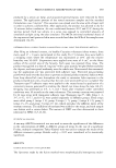
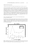
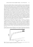
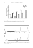
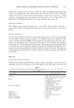
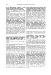
ANTI-INFLAMMATORY ACTIVITY OF C. ASIATICUM 423 µl of substrate (1 mM p-nitrophenyl-N-acetyl-13-D-glucosaminide) for 1 h at 37 ° C. Stop solution (0.1 M Na 2 CO 3 /NaHCO 3 ) was added, and the absorbance at 405 nm was measured with an ELISA reader. IN VIVO CLINICAL SAFETY TEST The evaluation of contact sensitization potential was carried out according to the meth ods previously reported by Shelanski and Shelanski (10). Fifty healthy Korean subjects were studied, and written consent was obtained in each case. The average age was 3 5 years (range: 21 to 51, males and females). The subjects had no skin disease, nor had they used topical or systemic irritant preparations in the previous month. All subjects were nonsmokers and signed an informed consent form in accordance with the Good Clinical Practice (GCP) and Declaration of Helsinki. Repeated epicutaneous applications under occlusive patch (repeat insult patch test, RIPT) for 48 h were performed using a Finn Chamber® (Epitest Ltd Oy, Finland) secured to the back site with Scanpore tape. These chambers are made of inflexible aluminum, and have a diameter of 12 mm and a depth of 0.5 mm. The round border of the chamber was placed firmly against the skin, causing a tight occlusion of the test materials. Sixty microliters of 1 % and 5% aqueous solutions of Crinum asiaticum extract were applied. The patches (chambers) stayed in place for 48 h for each application. The subjects abstained from showering or performing any work or exercise that might wet or loosen the patches. Eight applications were carried out for Cytotoxicity Assay 180 160 140 ;i' � 120 � 100 :Ei ftl 80 · "ii 60 40 Figure 1. Cytotoxicity of the ethanol extract of Crinum asiaticum to Raw 264.7 macrophage cells. N. con.: negative control (DMSO). P. con.: positive control treated with 1 µg/ml LPS. Indomethacin and the extract of Crinum asiaticum were treated with 1 µg/ml LPS. The data are expressed as the mean value (±standard deviations) of four experiments.
Purchased for the exclusive use of nofirst nolast (unknown)
From: SCC Media Library & Resource Center (library.scconline.org)