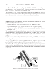
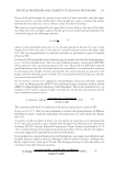
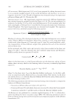
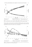
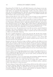
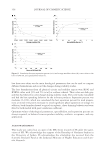
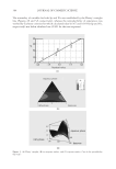
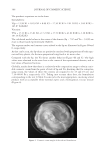
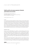
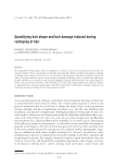
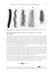
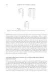
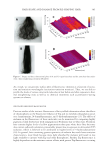
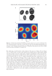
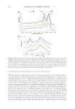
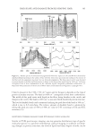
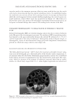
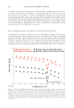
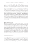
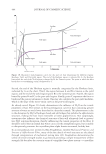
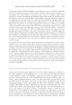
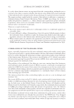
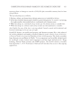
HAIR SHAPE AND DAMAGE FROM RE-SHAPING HAIR 387 THERMAL DEGRADATION OF HAIR Frequently, hair is exposed to thermal treatments to provide a desired hair set or style. In previous studies, we found that hair experiences surface (cuticular) and internal (cortical) damage as a result of thermal treatment (7). Hair also undergoes color changes on expo- sure to heat. This is clearly evident in the photograph of Piedmont hair shown in Figure 7, where one can visually observe the region of the tress where the hot iron treatment was administered, resulting in the formation of a dark yellow hue. In the case of dark brown hair, we do not observe a visually signifi cant color change, probably because it is masked by the absorption of melanin. Thermal treatment was administered for 1 min of Figure 6. Spectrofl uorescence excitation-emission matrix of Piedmont hair. Figure 7. Photographs of thermally exposed Piedmont and dark brown hair.

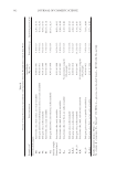
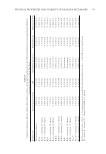
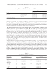
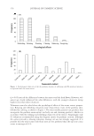
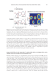
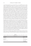
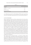
JOURNAL OF COSMETIC SCIENCE 388 continuous treatment. For this particular hot iron application, such a length of treatment might be considered extensive. However, this time scale was meant to be representative of cumulative treatments (i.e., a summation of a series of short treatment protocols) pro- viding an overall equivalent treatment time to the continuous treatment. In fact, previ- ous studies show that cumulative treatment is actually more damaging than continuous treatment (7). Data extracted from the excitation–emission matrices for thermally treated Piedmont and dark brown hair are provided in Table I. For dark brown hair, we observe a decrease in the residual Trp levels while the IKyn and I509 bands are statistically similar when com- paring the thermally exposed region of the tress with the unexposed portion. The calcu- lated peak ratios reveal the same information. In Piedmont hair, thermal exposure results in Trp loss, degradation of kynurenines, and an increase in the intensity at I509. As ex- pected, the ratio, ITrp/IKyn, decreases in thermally exposed hair as compared to the unex- posed region of the tress. In contrast, there is an increase in I509/IKyn in thermally exposed Piedmont hair. We suspect that the observed increase in this ratio is due to the formation of yellow coloration in thermally exposed hair. HAIR STRAIGHTENING The peak intensities and ratios for chemically relaxed hair are provided in Table II. As a result of the relaxer treatment, Trp fl uorescence decreases for both dark brown and Pied- mont hair (compared to untreated readings in Table I). In contrast, the signal for IKyn is essentially the same as for untreated hair in both dark brown and Piedmont hair. Al- though Trp degradation to kynurenines may occur—which would lead one to expect an increase in IKyn—the kynurenines themselves may be degraded by the relaxer treatment. In the case of the peak at the highest excitation wavelength employed, resulting in emis- sion at I509, relaxer treatment results in a large decrease in peak intensity for Piedmont hair and no change for dark brown hair. This effect may be more pronounced for Piedmont hair since its fl uorescence in this region is much more discernible. The peak ratios are also provided in Table II, which correspond with the peak intensity observations. INFRARED SPECTROSCOPIC IMAGING TO MONITOR LIPID DISTRIBUTION AND PROTEIN CONFORMATION IN HAIR Infrared (IR) spectroscopy is an indispensable tool for the study of biological samples due to the inherent chemical specifi city of vibrational frequencies in the IR spectrum. By coupling an FT-IR spectrometer with a microscope, one may perform FT-IR spectro- scopic imaging, which allows for the measurement of the chemical environment of a specifi c area of a specimen. In this way, we can spatially resolve molecular structure infor- mation. In essence, a two-dimensional matrix, composed of pixels, contains an IR spec- trum in each pixel. This allows us to generate FT-IR images corresponding to a chosen vibration frequency of interest. In the past two decades, FT-IR spectroscopic imaging has transformed into a powerful biophysical approach to study various types of biological tissues (18–21). The spatially resolved spectroscopic images generated from this technique provide a histological map
Purchased for the exclusive use of nofirst nolast (unknown)
From: SCC Media Library & Resource Center (library.scconline.org)