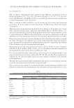
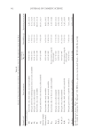
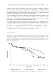
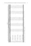
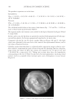
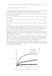
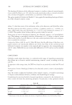
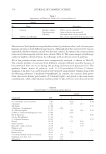
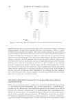
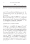
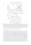
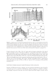
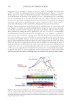
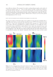
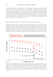
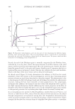
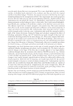
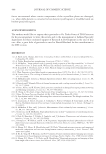
HAIR SHAPE AND DAMAGE FROM RE-SHAPING HAIR 407 Table IV Tensile Strength Data for Chemically Treated Hair Measured in the Fully Hydrated State (48) Hair sample Tensile strength (N·m)/1000) Untreated 1.21 Hair color (30 vol. developer) 1.14 Acid wave 0.99 Hair relaxer (NaOH) 0.78 Hair relaxer (guanidine) 0.70 Hair bleach (30 vol. developer) 0.48 even a slight increase in some of the tensile parameters. Similar to the case of wool, heat- ing to elevated temperatures may result in the formation of cross-links within the inter- nal structure of hair thereby requiring greater forces to extend and break the fi ber (50). CONCLUDING REMARKS Chemical and physical treatments intended to change the shape of hair often result in changes to fi ber shape, which is often accompanied by damage to structural proteins of the fi ber. Photographic imaging techniques in conjunction with image analysis provide accurate measurements of fi ber shape as manifested in fi ber alignment and curl. In addi- tion, using laser stereometry we characterize the three-dimensional structure of the fi ber assembly and its occupied volume, allowing for comparison of the initial state with that of the reshaped hair. On the other hand, many other tools are available to assist in deter- mining the location and extent of damage associated with reshaping hair. Spectroscopic techniques are the best tools at providing us with real chemical information as to the health state of hair. These changes can be probed by monitoring specifi c amino acid resi- dues utilizing fl uorescence spectroscopy, Raman spectroscopic imaging, or IR spectro- scopic imaging. As an example, one may monitor tryptophan degradation with fl uorescence, or conversion of cystine to cysteic acid residues by IR spectroscopic imag- ing. In all of the IR imaging data presented, we examined cross sections of hair fi bers, providing specifi c chemical information in a spatially resolved manner. All of the spectro- scopic techniques are advantageous however, Raman confocal imaging is extremely pow- erful since it permits us to noninvasively dissect the fi ber providing a z-line (through the fi ber cross section) of functional group or secondary structure information. Both spectro- scopic imaging methods also allow for the determination of beta keratin, and its local- ization in the morphological components of hair. In this way, we monitored the conversion of alpha keratin to beta keratin. More common techniques to measure hair damage—especially due to perming, straight- ening, and fl at ironing—include DSC and tensile property measurements. The increased utility of these techniques stems from their ease of use and instrument accessibility (e.g., budgetary factors). DSC provides us with a quick look at the health state of the crystalline (alpha-helical component) and amorphous (the matrix located in cortical cells) regions of hair by monitoring ΔHD and TD. Tensile strength measurements, on the other hand, are normally used to generate data about the tensile properties of hair. These data are typically presented in the form of stress–strain curves that provide a fi ngerprint of the

JOURNAL OF COSMETIC SCIENCE 408 forces encountered when certain components of the crystalline phase are damaged i.e., when alpha keratin is converted to beta keratin (yield region) or disulfi de bonds are broken (postyield region). ACKNOWLEDGEMENTS The authors would like to express their gratitude to Dr. Trefor Evans of TRI-Princeton for his encouragement to write this article and to the management at Ashland Specialty Ingredients for their continued support of Research & Development in the area of hair care. Also, a great debt of gratitude is owed to Donald Koelmel for his contributions to the DSC section. REFERENCES (1) A. Byrd and L. Tharps, Hair Story: Untangling the Roots of Black Hair in America (Diane Pub Co., Collingdale, PA, 2006). (2) A. Valdez, Brazilian hair straightening, Cosmetiscope, 17(6), 1–7 (2011). (3) R. McMullen, “Image analysis tools to quantify visual properties of hair fi ber assemblies,” in Practical Modern Hair Science, T. Evans and R. Wickett. Eds. (Allured: Carol Stream, IL, 2012), pp. 295–332. (4) R. Asquith M. S. Otterburn, and J. A. Swift, Physical changes occurring in heated mammalian keratin, J. Text. Inst., 63, 544–550 (1972). (5) I. Watt, Properties of wool fi bers heated to temperatures above 100°C, Text. Res. J., 45, 728–735 (1975). (6) M. Gamez-Garcia, The cracking of human hair cuticles by cyclical thermal stresses, J. Cosmet. Sci., 49, 141–153 (1998). (7) R. McMullen and J. Jachowicz, Thermal degradation of hair. I. Effect of curling irons, J. Cosmet. Sci., 49, 223–244 (1998). (8) S. Ruetsch and Y. Kamath, Effects of thermal treatments with a curling iron on hair fi ber, J. Cosmet. Sci., 55, 13–27 (2004). (9) T. Inoue, M. Ito, and K. Kizawa, Labile proteins accumulated in damaged hair upon permanent waving and bleaching treatments, J. Cosmet. Sci., 53, 337–344 (2002). (10) M. Wong, G. Wis-Surel, J. Epps, Mechanism of hair straightening, J. Cosmet. Sci., 45, 347-352 (1994). (11) J. Russ, The Image Processing Handbook, 4th Ed. (CRC Press: Boca Raton, FL, 2002). (12) B. Pourdeyhimi and H. S. Kim, Measuring fi ber orientation in nonwovens: The Hough transform, Text. Res. J., 72, 803–809 (2002). (13) G. Loussouarn, A. Garcel, I. Lozano, C. Collaudin, C. Porter, S. Panhard, D. Saint-Léger, and R. de La Mettrie, Worldwide diversity of hair curliness: A new method of assessment, Int. J. Dermat., 46 (Suppl. 1), 2–6 (2007). (14) R. McMullen, F. Zisa, and J. Jachowicz, Measurements of hair volume by laser stereometry, J. Cosmet. Sci., 60, 171–185 (2009). (15) J. Jachowicz and R. McMullen, Tryptophan fl uorescence in hair—examination of contributing factors, J. Cosmet. Sci., 62, 291–304 (2011). (16) S. Daly, R. Bianchini, T. Polefka, L. Jumbelic, and J. Jachowicz, Fluorescence and coloration of grey hair, Int. J. Cosmet. Sci., 31, 347–359 (2009). (17) J. Prompers C. W. Hilbers, and H. A. M. Pepermans, Tryptophan mediated photoreduction of disul- phide bond causes unusual fl uorescence behavior of Fusarium Solani pisi cutinase, FEBS Lett., 45, 409– 416 (1999). (18) G. Zhang, D. J. Moore, R. Mendelsohn, and C.R. Flach, Vibrational microspectroscopy and imaging of molecular composition and structure during human corneocyte maturation, J. Invest. Dermatol., 126, 1088–1094 (2006). (19) X. Bi, X. Yang, M. P. Bostrom, and N. P. Camacho, Fourier transform infrared imaging spectroscopy investigations in the pathogenesis and repair of cartilage, Biochim. Biophys. Acta, 1758, 934–941 (2006). (20) G. Zhang, D. J. Moore, C. R. Flach, and R. Mendelsohn, Vibrational microscopy and imaging of skin: from single cells to intact tissue, Anal. Bioanal. Chem., 387, 1591–1599 (2007).
Purchased for the exclusive use of nofirst nolast (unknown)
From: SCC Media Library & Resource Center (library.scconline.org)