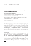
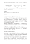
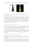
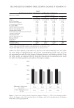
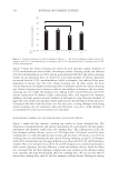
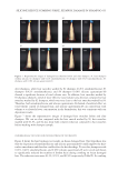
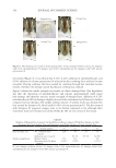
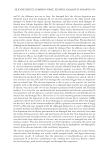
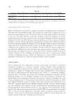
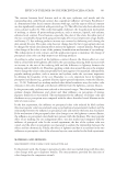
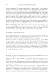
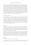
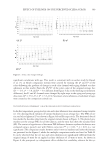
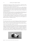
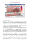
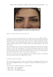
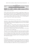
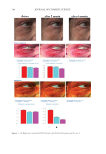
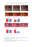
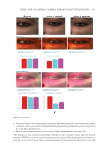
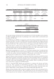
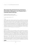
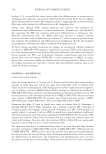
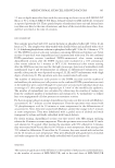
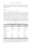
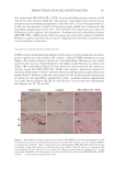
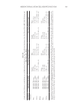
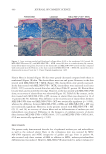
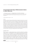
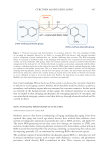
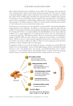
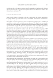
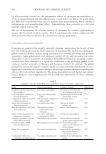
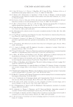
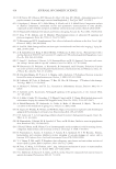
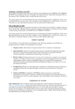
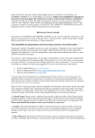
380 JOURNAL OF COSMETIC SCIENCE OBJECTIVES AND METHODS Included criteria. The study included 50 adult, males and female, aged 22–55, with infraorbital dark circles they were recruited from a dermatology outpatients’ clinic. Only 40 patients were included in the statistics, as 10 patients discontinued treatment and were excluded from the study. Other exclusion criteria. These included pregnancy and lactation, skin malignancies, autoimmune diseases, thrombocytopenia, platelet dysfunction syndrome, and anticoagulant drugs and previous treatments for infraorbital region in the last year. All included patients were subjected to detailed personal, present, family, and drug history recording. Dermatological examinations were done, including photographic documentation of lesions before and after 1 and 6 months of treatment. Treatment response was assessed using an Antera 3D® (Miravex Limited, Dublin, Ireland) camera before and after treatment. The treatment area was the infraorbital region, extending from the lower upper eyelid superiorly and the inferior orbital rim inferiorly. The medial canthus angle (medially) and the lateral canthus angle (laterally) are shown in Figure 4 (10). Treatment procedures. The technique of preparing plasma gel was described by Gupta et al. They mentioned that plasma serum was centrifuged and calcium gluconate was added in the platelet-poor plasma (PPP) in the proportion of 0.01 mL/1 mL of PPP. It was incubated in hot water at 100°C for 5 minutes and then in a cold-water bath in a metal container, with an initial temperature of 7 °C, for 5 minutes. Then, the gel was ready (11). In this study, the technique was modified to a novel one. A total of 10 mL of the patient’s blood was taken and mixed with 1 mL of anticoagulant acid citrate dextrose. It was rotated at 3,000 rotations per minute for 10 minutes in a centrifugator as a single spin. PPP and platelet-rich plasma (PRP) were withdrawn in a 1-mL syringe, and approximately 5 Figure 3. Haemoglobin mode in Antera camera (9).

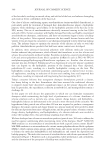
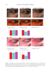
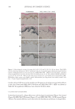
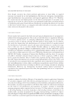
381 Using the 3D Antera Camera for Accurate Evaluation syringes were extracted. Calcium gluconate was added to the mixture of PPP +PRP in a concentration of 0.01 mL/1 mL. The mixture was then placed into a plasma filler machine and heated to 90 °C (5 minutes), then cooled to 70 °C (5 minutes), and finally cooled to 40 °C (5 minutes). The viscous gel was then ready for use. Topical anesthetic cream was applied over the treated areas for 45 minutes. The gel was injected using a 1-mL, 4-mm 27 G needle. The left infraorbital area was injected with plasma gel (filler) in the deep dermis as multiple injections, with 0.1 mL in each one, 0.5 cm apart. This procedure was done for three sessions, 1 month apart. The PDO mono threads (Venus V Line®, TM 25 mm, Thread 30G, Korea) were inserted in the right infraorbital area for one session. The numbers of threads were between three and five, inserted in the deep dermis. Post-treatment. A sunscreen cream was applied after each treatment session to be used daily before sun exposure and to be reapplied every 2 hours. STATISTICAL ANALYSIS OF THE DATA The collected data was revised, coded, and tabulated using Statistical Package for the Social Sciences (IBM SPSS Statistics for Windows, Armonk, NY: IBM Corp.,Version 25.0. Armonk). The p value was considered significant as the following: • The p value 0.05: nonsignificant. • The p value 0.05: significant. • The p value 0.01: highly significant. Figure 4. Anatomical landmarks of periorbital region (10).
Purchased for the exclusive use of nofirst nolast (unknown)
From: SCC Media Library & Resource Center (library.scconline.org)