J. Soc. Cosmet. Chem., 21,583-594 (August 19, 1970) Chemical Differentiation in the Epidermis* I. A. BERNSTEIN, Ph.D.* Presented December 2, 1969, New York City Synopsis--In its progress from the basal layer toward the outer surface of the skin, the EPIDERMAL CELL undergoes a series of morphological changes .which undoubtedly coincide with alterations in metabolic activity. Study of these biochemical modulations should provide an understanding of the mechanisms basic to the mediation and control of epidermal differentiation culminating in KERATINIZATION, The exclusive localization of an unusual PROTEIN in the cells which contain the keratohyalin granules provides a vehicle for such an investigation. The rapid synthesis and the location of the protein in the keratohyalin-containing cells underlying the keratinized layer, the presence of a large amount of histidine in the keratohyalin, and the absence of the "histidine-protein" when keratohyalin is absent in psoriasis suggest an association of the protein and keratohyalin and support the hypothesis that both are involved in epidermal keratinization. Availability of an in vitro system for the synthesis of this protein would allow a study of the mechanisms of control of CHEMICAL DIFFERENTIATION which the epidermis can invoke. INTRODUCTION The mammalian cutaneous epidermis plays a significant role in maintaining a stable internal environment for the organism. While carrying out this protective function, the tissue is continuously being tormented by chemical, physical, and biological agents in the external * Work supported by Grants AM 10225 and AM 05268 from the National Institutes of Health, United States Public Health Service. • Cellular Chemistry Laboratory of the Departments of Dermatology and Environmental and Industrial Health, and the Department of Biological Chemistry, The University of Michigan Medical Center, Ann Arbor, Mich. 48104. , 583

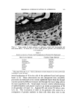
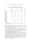
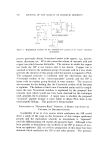
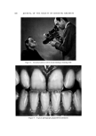
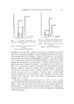
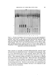
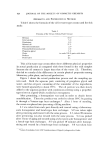
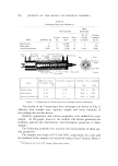
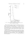
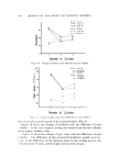
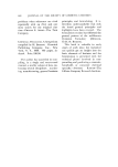
584 JOURNAL OF THE SOCIETY OF COSMETIC CHEMISTS environment. Ordinarily, the normal rate and sequence of epidermal cellular renewal adequately counteracts the trauma of external environ- mental stress. A detailed knowledge of the molecular mechanisms in- volved in epidermal differentiation should provide a rational basis for the design of therapeutic and cosmetic measures and agents to assist the epidermis when, for any reason, its proliferative ability is inadequate for preserving its integrity and function. Current attempts to define the nature of specific epidermal proteins and to localize their biosynthesis in particular stages of morphological differentiation are a beginning to- ward elucidating the mechanisms controlling the ordered sequence of structural changes which culminates in the formation of a cornified outer layer in the skin. Control mechanisms of epidermal differentiation would seem to necessarily involve the genetic apparatus. Presumably, epidermal DNA contains operational instructions and information on the chronolo•oy of events in the process of differentiation. It is possible that the environ- ment might be involved in initiating the specific steps (i.e., in modulating the timing of the events), but the consistency in the sequence of steps ap- pears to require an inherent programming of the process with the en- vironment playing only a modifying role. The mechanisms of molecular change and chronological control of the steps could be studied if one of the molecular events could be defined and could be localized in the sequence, preferably in relation to a morphologically recognized change. The synthesis of a unique protein in the •oxanular cell of the newborn rat epidermis appears to provide a vehicle for such a study and this report will review the data developed in this laboratory relating to this biochemical system. I,OCI OF EPIDERMAL PROTEIN SYNTHESI •, Figure 1 is a photomicrograph of the epidermis of the newborn rat illustrating the single layer of basal cells at the dermal-epidermal junction, the several layers of larger spinous cells lying above the basal layer, and the thick zone of cells containing keratohyalin granules just under the cornified outer stratum. Normally, mitosis is restricted to the basal layer with, statistically, one of the two daughter cells leaving this germina- tive layer to enter the sequence of differentiation ultimately being sloughed from the skin surface as a fully keratinized cell. As shown in Table I, the intraperitoneal injection of tritiated4eucine, valine, methionine, and phenylalanine to newborn rats results in an
Purchased for the exclusive use of nofirst nolast (unknown)
From: SCC Media Library & Resource Center (library.scconline.org)