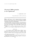
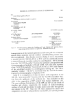
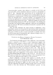
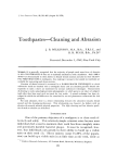
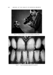
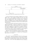
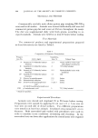
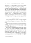
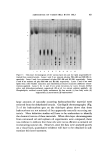
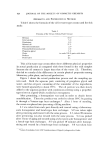
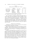
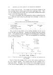
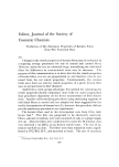
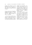
CHEMICAL DIFFERENTIATION IN EPIDERMIS 585 • • .. . •. y .. . • • .. • . ..-•. •-,•,:• ,.......,. ..•: . :-...: :.:,:,•:.- • •,--: . . . .. •.-:"..:.,,•.., .•?• - • ]•. '•'• . •.• . . . ..:. ... • ..... . • .•: ... . . .%-• .., . :- ..... :-:3 ...... ....• ....,.. ..... - ..• .•. •..•,•,.4• • --•,. . .... ...... • .... •. •... .•g . .•, . ,?ff ' .-• ß ':'•'.' /•g:'d::•'. " , •- . ß .•' ... ',•:•'.' . '• : •: . ,... .• .... ..... • ..•... ---]:'• . : -- • . ,. w •- :. ' -" • - •- ."•, •' ' 2 . •.'• -.:-, ...... ." -k •: : - •, '•.•-'•' ' • . •. :•? ---• : .... ' •*•?..- -•- .,--•'•%. ,,'•:2 ,...-%•% •. *-- 'a ..... . ......... ...':• '•" .k:.'.- •..' • ß ..•'•'•" -•. :•2 ........... ß •:? -:: ' '•..•' •.•[:- -•.•?•---- :, •: . • .. •:--•- .•.. .• • .. • . .• ••. -•- ..•. :• • .• Figure 1. Tissue section of whole newborn rat epide•is stained with hematoxylin and eosin (about 600X). D--denis B, S, G, and C--baml, spinous, granular, and cornified la•'ets of the epidermis Table I Initial Localization of Amino Acid Incorporation in Newborn Rat Epidermis• Basal (Lower Spinous) Granular (Upper Spinous) Leucine Histidine Methionine Glycine Phenylalanine Arginine Lysine Serine Tyrosine Tyrosine Proline Proline Valine • Data taken from refs. (1, 2). Amino acids shown in both columns do not show preferential localization in any cell layer. initial localization of aH in the cells of the epidermal basal and spinous layers (1, 2). These observations are not unexpected since metabolic precursors are delivered to the various epidermal layers from the dermal vascular system by diffusion between and through the lower lying cells to the more superficial cells. Surprisingly, however, similar administration of labeled histidine and glycine results in an initial accumulation of aH in the granular cell layer (1). Arginine-aH and serine-aH behave simi- larly (2). This localization of label in the granular cells cannot be a consequence of protein synthesis in these cells when they were in the

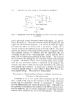
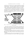
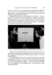
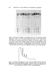
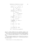
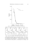
586 JOURNAL OF THE SOCIETY OF COSMETIC CHEMISTS basal or lower spinous layers, since the initial accumulation of histi- dine-all in the granular layer is seen by 15 minutes after the intra- peritoneal injection of the tracer (1) and it takes of the order ot! three days for a cell to move from the basal into the •anular layer in the epidermis ot5 the newborn rat (3). A similar differential localization oc- curs in human epidermis (4, 5). A variety of possible explanations could be proposed for this unusual localization of certain amino acids in the gTanular layer (6). The most simple and provocative hypothesis would be to propose that there is a rapid biosynthesis ot5 proteins in these cells--proteins which have uniquely high levels of these amino acids and little or no concentration of the amino acids initially incorporated into the lower cells. Since RNA can be synthesized in the granular cells (7) even though DNA is not (3), protein synthesis in these cells is a tenable hypothesis. The epidermis of the newborn rat does indeed contain two species of protein with high levels of histidine and glycine, respectively, which become labeled rapidly (6). Neither contains significant amounts of phenylalanine, leucine, valine, or inethionine. Furthermore, the "histi- dine-rich" protein (HP) has been localized in the cells of the granular layer (8). Cysteine residues in the free sulfhydryl or disulfide form are not detected in either the HP or "glycine-rich" protein (GP) in spite of lhe preferential initial incorporation of cystine-aH in the granular cells (9). Fractionation of epidermal protein from an animal which had re- ceived cystine-a or cystine-"S intraperitoneally indicates clearly that this amino acid is incorporated in the granular layer into proteins which are different from those containing the high level of glycine and histi- dine (10). Although it appears from kinetic studies of amino acid incorporation visualized by autoradiography (1) that protein synthesis occurs in all vi- able cells of the epidermis of the newborn rat, two major loci ot5 protein formation exist. One is in the basal and lower spinous cells while the other is in the granular cells. Protein synthesis in the latter locus probably leads to the formation of the unique HP and GP and a protein with a high concentration of cysteine residues, yet to be isolated. NATURE OF THE "HISTIDINE-RICtI" PROTEIN Figure 2 is a flow diagram of the technique, slightly modified from that previously published (6), which is used to obtain the HP and GP froxn the epidermis of the newborn rat. The extract resulting from
Purchased for the exclusive use of nofirst nolast (unknown)
From: SCC Media Library & Resource Center (library.scconline.org)