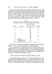
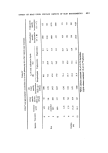
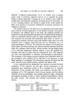
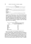
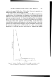
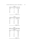
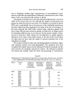
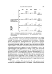
488 JOURNAL OF THE SOCIETY OF COSMETIC CHEMISTS of the treatment group. Epidermal cells were harvested from the pooled sections by mild trypsinization (1). These were then cultured with •4C- glycerol. Samples of the epidermal sections were also cultured. After cell and tissue cultures, lipid radioactivities and total DNA levels were measured. Radioactive lipids were analysed by thin-layer chromatography, the results of which are shown in Table VL Table VI Glycerolipid metabolism in whole epidermis and isolated epidermal cells of soap-treated rat skin* Group Tissue Lipid Lipid labelling pattern specific (% of total lipid radioactivity) incorporation (dpm pig -x DNA) Phospholipids Triglycerides Diglycerides Control Whole epidermis Isolated cells Soap Whole treated,' epidermis Isolated cells 1 702 85.6 11.8 2.7 747 76.3 17.4 6.3 1 847 74.9 25.0 0.0 1 029 48.7 48.2 3.1 *Tissues cultured with 5.0 •Ci U-x4C-glycerol. •-8 % of soap formulation in water. pH of solution ----- 9.8. In the control rats it was seen that the specific incorporation into the lipids of the whole epidermis was far higher than that of the isolated epidermal cells, and suggested that there were other regions of the epidermal sample with significant lipogenic activity too. The distributions of •4C- glycerol between various glycerolipids were quite similar, however, with most radioactivity found in phospholipids. After soap treatment, however, certain changes were seen. The specific incorporation into lipids of the whole epidermis increased by less than 10•o, whereas in the isolated epidermal cells this parameter increased by nearly 40•o after soap treatment, and indicated that the stimulated lipid synthesis was concentrated in the Malpighian cells of the epidermis. Similarly, the distribution of radioactivity among different glycerolipids was altered in soap-irritated tissues. In the cultured whole epidermal sections the proportion of labelled phospholipids dropped from 85.6 to 74.9•o of the total lipid radioactivity, with a concomitant rise in triglyceride radioactivity, a feature already amply illustrated in the experiments above. However, in isolated cells of

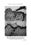
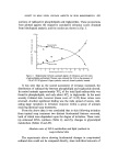
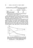
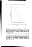
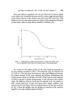
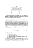
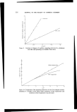
EFFECT OF SOAP UPON CERTAIN ASPECTS OF SKIN BIOCHEMISTRY 489 the epidermis these changes were far more prominent. The proportion of labelled phospholipids dropped from 76.3 to 48.7•o, and labelled tri- glyceride rose from 17.4 to 48.2•o. This amplification of the soap-induced changes in lipogenesis within the isolated Malpighian cells, compared with whole epidermis suggested, therefore, that the Malpighian layer was the site of altered metabolic activity. DISCUSSION The effects of soaps on mammalian skin have been amply documented since the first observations by Emery and Edwards (3). More recently Bettley (4) reviewed soap action in terms of its antibacterial action, skin cleansing ability, effect on water permeability and effect on the stability of the acid mantle of skin. Wood and Bettley (5) described changes in the levels of liberated sulphydryl group in human keratin after denaturing with soap solutions. Emery and Edwards (3) showed in an homologous series of fatty acids that sodium laurate exhibited the most frequent irritation on human skin. Choman (6), in an in vitro test, showed that sodium laurate caused far greater swelling of collagen than other soaps. These findings have been corroborated by Fig. 2 where topically applied sodium laurate not only caused much greater irritation than other constituent fatty acids of the soap, but also induced the most marked changes in biochemistry (Fig. 5). From this we conclude that both the irritation potential of the soap and the re- sultant degree of biochemical alterations induced are due principally to its lauric acid content. The findings of this study may be summarized as follows. When rats are treated topically with soap solutions an irritation response develops which is generally proportional in severity to the number and frequency of treatments. At the microscopic level this irritation is seen as an increase in the proliferative processes of the epidermis, seen as a thickened Malpighian layer, a thickened, compacted and parakeratotic stratum corneum, contain- ing nuclear debris, and with invasion of leucocytes from the underlying dermis. In association with these morphological changes we have observed a stimulated DNA synthesis and an altered glycerolipid metabolism. No distinct sequence of these biochemical phenomena has been established, although from Fig. 4 and Table IV it appears that DNA metabolism in- creases early in the response of the tissue to soap treatment, and is pre- requisite for the entry of more cells of the basal layer into mitosis. A feature of the DNA specific activities was that after reaching a maximum
Purchased for the exclusive use of nofirst nolast (unknown)
From: SCC Media Library & Resource Center (library.scconline.org)