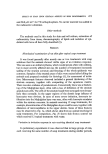
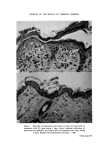
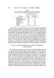
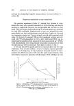
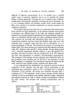
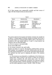
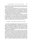
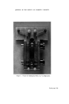
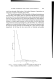
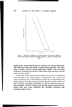
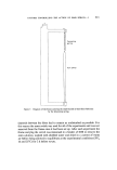
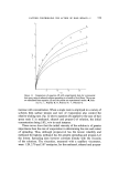
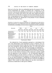
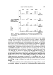
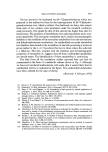
EFFECT OF SOAP UPON CERTAIN ASPECTS OF SKIN BIOCHEMISTRY 475 and 50.0 [tCi ml -• for a2P-orthophosphate. No carrier material was added to the radioactive substrates. Other methods The methods used in this study for skin and cell culture, extraction of radioactivity from tissue, chromatography of lipids and isolation of epi- dermal cells have all been fully described (1). RESULTS Histological examination of rat skin after topical soap treatment It was found generally after merely one or two treatments with soap solutions that the animals showed visible signs of an irritation response. This was seen as an initial erythema, which would persist often with oedema, but in more severely affected skin (as the number of treatments increased) scaling of the stratum corneum and fissuring of the whole epidermis was common. Samples of the treated areas of skin were excised after killing the animals and prepared routinely for histology (1), for assessment of irrita- tion. Microscopic features observed included a general thickening of the stratum corneum, together with compacting of the squamous matrix. There was also evidence of parakeratosis. Concurrently there was a thicken- ing of the Malpighian layer, often with a loss of definition of the stratum granulosum cells. The cells of the stratum basale were arranged more irregu- larly than normally. In the upper regions of the dermis large influxes of leucocytes were obvious. In cases of more severe irritation, leucocyte in- filtration into the Malpighian layer was seen, often with leucocyte exudates within the stratum corneum. In animals receiving 12 soap treatments, for example, discontinuities of the Malpighian layer could be seen, together with dilatation of microcapillaries in the upper dermis. Some of these features may be observed in Fig. la, which shows skin from an animal receiving 12 soap treatments. For comparison, Fig. lb shows skin from a control rat which received 12 topical treatments with water. Variation in irritation response in rats receiving identical soap treatments In preliminary experiments it was observed that in large groups of rats, each receiving the same number of soap treatments during similar periods,

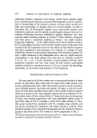
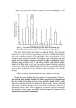
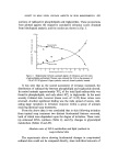
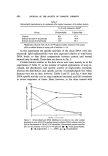
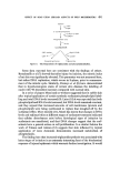
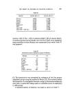
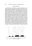
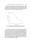
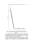
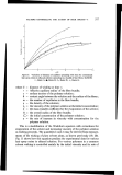
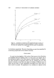
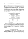
476 JOURNAL OF THE SOCIETY OF COSMETIC CHEMISTS individual irritation responses were always varied. Some animals might show well-developed responses as measured histologically in terms of altera- tions in morphology of the stratum corneum, whereas others would show little, and occasionally, no changes relative to control animals. In order to rationalize this, all histological samples from soap-treated animals were subjectively analysed, and the specific morphological changes observed (i.e. corneum thickening, leucocyte infiltration, capillary dilatation etc.) were assessed under increasing degrees of severity.* These arbitrary categories were then given a numerical weighting as follows: very slight reaction (numerical weighting = 0.25) slight reaction (0.5) fairly distinct reaction (0.75) quite distinct reaction (1.0) well-developed reaction (2.0) and severe reaction (3.0). The cumulative score for the criteria of the irritation response were then compiled for each specimen. For example, if a sample of soap- treated skin showed a quite distinct thickening of the stratum corneum, but only a slight thickening of the Malpighian layer, and very slight leucocyte accumulation in the upper dermis, the total score of irritation would be 1.75 (i.e. 1.0 + 0.5 + 0.25). Generally, control animals receiving water treatments exhibited very low total scores (0-2.0) whereas soap-treated animals would have cumulative scores of 3.0 up to maybe 20, depending, necessarily, on the number and duration of the treatments. Individual fatty acids and irritancy of skin The soap used in all of these studies was a conventional mixture of palm kernel oil and tallow fatty acids and the major fatty acids present were laurate, palmirate and oleate. Other fatty acids, present in lower concentra- tions included stearate, myristate and caprate. In order to test the contri- bution of individual ingredient fatty acids to the overall irritation response observed, a group of rats were topically treated with pure solutions of cer- tain sodium soaps of fatty acids as follows. Twenty-seven rats, in nine groups of three, were treated respectively with seven individual sodium soaps of pure fatty acids, at 0.25 M strength, a total of five times during 21/: days. One group received five treatments with 8•o complete soap solution, and one group, receiving five treatments with water, served as controls. After treatment samples were examined histologically and the irritation responses scored as described above. The results are shown graphically in Fig. 2. *The authors acknowledge the help and criticism given by Mr H. L. Jenkins, who also developed the method for quantitative histological assessment of irritation.
Purchased for the exclusive use of nofirst nolast (unknown)
From: SCC Media Library & Resource Center (library.scconline.org)