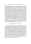
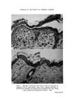
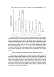
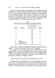
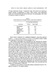
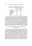
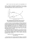
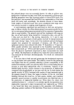
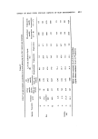
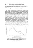
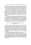
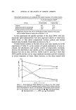
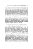
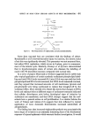
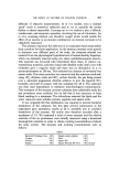
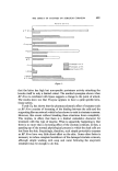
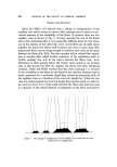
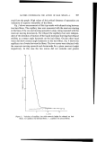
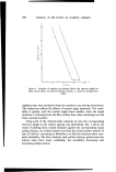
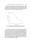
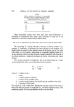
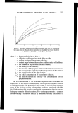
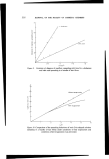
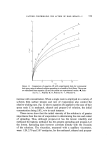
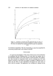
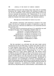
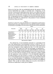
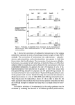
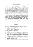
490 JOURNAL OF THE SOCIETY OF COSMETIC CHEMISTS they declined with increasing treatments. In Fig. 5 in particular, where skin had undergone severe treatment with sodium Iaurate DNA metabolism was virtually zero after eight applications, this probably being due to the com- plete necrosis of the cells of the germinative layer of the epidermis, which would be unable to perform anabolic reactions. The a•'P-orthophosphate experiment of Fig. 4 and the xaC-glycerol experiment of Table IV show that in rat skin treated with soap, phospholipid radioactivity also reached a maximum, similar to DNA data. This parallel may indicate an initial hyper- trophy in the epidermal cells, which would precede the hyperplasia. seen histologically, at later stages of the irritation response. In a similar fashion to DNA labelling, specific incorporations into phos- pholipid also generally decreased after prolonged treatment. This, again, may have been due to general impairment of the tissue's ability to supply the cyti- dine cofactors required for phospholipid synthesis after more severe irritation. The most novel finding, however, was the dramatic increase in trigly- ceride labelling in soap-irritated skin. This lipid is not a classical membrane component, as are phospholipids and sterols, but is generally agreed to serve as an energy store. The proportion of radioactive triglyceride in the total labelled lipids rose as the irritation response developed (Fig. 2), and occurred much later in the time scale of the irritation response than the stimulation of DNA synthesis. Fig. 5 is remarkable in that it shows the massive tri- glyceride synthesis in sodium laurate-treated skin, in which DNA and phospholipid synthesis had virtually ceased. In this experiment the overall level of total lipid synthesis did not diminish greatly with soap treatment but thin-layer analyses showed that the high level of phospholipid synthesis of the control was largely replaced by an almost equally high level of tri- glyceride synthesis. There was no obvious stoichiometric relationship between these two lipid types, because in Table IV, for both the rat and the guinea-pig total lipid specific incorporation greatly exceeded the control levels (Table IV). Fig. 6 shows the biosynthetic pathway for certain glycerolipids, and illus- trates that both triglycerides and phospholipids share a common precursor, namely, 1,2-diglyceride. One might argue that a block in phosphatidyl- choline synthesis at the CDP-choline formation steps could lead to an ac- cumulation of 1,2-diglyceride, which could then be further acylated to tri- glyceride. However, this would not explain why the total amount of lipid radioactivity ix mole -x of DNA was consistently higher in soap-treated skin compared with controls.

EFFECT OF SOAP UPON CERTAIN ASPECTS OF SKIN BIOCHEMISTRY 491 Glycerol I .•--• AT P •'ADP 25-G!ycerophosphate I •2 AcyI-S-CoA •'- CoASH Phosphatidic acid 1,2-Diglyceride CDP-choline. / • _..Acyl-S-CoA CMP '""•• '• CoASH Phosph•tidylcholine Triglyceride Figure 6. The biosynthesis of triglycerides and phosphatidylcholine. Some data reported here are consistent with the findings of others. Bertalanaffy et al (7) showed that after injury by incision, the mitotic index of rat skin was significantly elevated. This parameter was not measured here, but rather DNA replication, which occurs in S-phase, prior to commence- ment of the mitotic cycle. Similarly, Penneys et al (8) have demonstrated that in hyperkeratogenic states of certain skin diseases the labelling of nuclei with aH-thymidine increases compared with normal skin. In a series of papers Mezei and co-workers suggested that in rabbit skin after topical application of certain synthetic surfactants phospholipid label- ling and total DNA levels increased (9). Later (10) it was reported that both phospholipid and RNA levels increased, but DNA levels remained constant, and they argued that increased amounts of cell membranes (protein and phospholipid) were being synthesized to replace that sloughed off by the surfactant effect. More recently (11), Mezei has shown that changes in RNA levels and radioactivities at different stages of surfactant treatment indicated that cellular disturbances exist before histological signs of irritation by surfactants are manifested, and that DNA changes suggest that the surf- actants have a general action on cell proliferation. In a similar fashion the work of Takasu and Aizawa (12) suggests that skin inflamed by topical application of toxic chemicals demonstrates increased metabolism of phospholipids. The finding here that increased triglyceride synthesis was associated with latter stages of irritation is an extremely interesting facet of the biochemical response of injured epidermis which warrants further investigation. It would
Purchased for the exclusive use of nofirst nolast (unknown)
From: SCC Media Library & Resource Center (library.scconline.org)