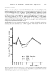
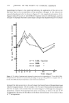
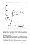
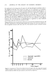
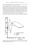
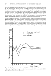
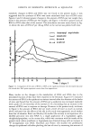
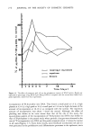
j. Soc. Cosmet. Chem., 30, 263-278 (September/October 1979) Some biochemical effects of isopropyl myristate and squalane on rabbit skin H. KOMATSU, K. ASABA and M. SUZUKI Pola Laboratories, 27-1, Takashimadah Kanagawa- ku, Yokohama, Japan 221. Received December 13, 1978. Presented at the loth IFSCC Congress, October 1978, Sydney, Australia. Synopsis A biochemical method was employed to study the responses of RABBIT SKIN to ISOPROPYL MYRISTATE, SQUALANE, and DECANE. Skin samples excised with a Castroviejo keratotome were separated into lipid, TCA-soluble, RNA and DNA fractions after incubation with 40 •tCi Na2H32PO4 at 37øC for 1 hr. BIOCHEMICAL CHANGES in the epidermis with time were evaluated in terms of the changes in total phosphorus contents, specific activity and incorporated amount per unit DNA of each fraction. The results showed that decane damaged the skin so severely that the biosyntheses of lipids, RNA and DNA were reduced markedly for the first three days after application, but increased rapidly after that due to the repair. The effect of squalane was found to be weaker than that of isopropyl myristate, though both oils induced the stimulation of biosyntheses in the epidermis. The magnitude of the biochemical effects of the three oils on the skin was increased in the order of squalane, isopropyl myristate and decane, which was consistent with the results of macroscopic and histological observations. From the profiles of the effects it is postulated that the repairing processes are controlled by some feedback mechanisms. INTRODUCTION There are many studies on the biochemical changes in the skin caused by the percutaneous application of a chemical substance. Aso and Okazaki (1) measured the epidermal mitotic activity with 3H-thymidine and the biosynthesis of epidermal lipids with •4C-malonyl CoA and •4C-mevalonic acid after the application of vitamin A acid on human psoriatic skin and normal guinea pig skin. Prottey et al. (2) studied the effects of soaps upon the metabolism of DNA and lipids of rat skin with the aid of •4C-thymidine, 32p:orthophosphate, •4C-acetate and •4C-glycerol. Mezei (3) investigated the effect of polysorbate 85 upon human skin in terms of the changes in the rate of 32p incorporation into lipid, TCA-soluble, RNA and DNA fractions after incubating the skin sample in a medium with Na2H32PO4 . Stegman et al. (4) observed the inhibition of the effect of concanavalin-A on the epidermis of newborn rats by o•-methyl- D-glucopiranoside in terms of the incorporation of 3H-thymidine and 3H-histidine. On the other hand, there are few reports on the effects of the oils commonly employed 263

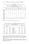
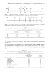
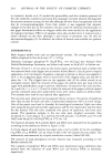
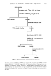
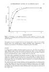
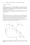
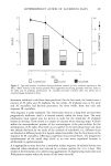
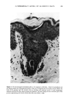
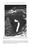
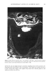
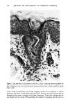
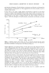
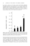
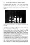
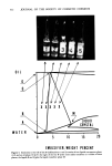
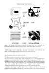
264 JOURNAL OF THE SOCIETY OF COSMETIC CHEMISTS in cosmetics. Suzuki et al. (5) studied the permeability and skin irritation potential of five oils useful for cosmetics and found that isopropyl myristate showed histologically the severest irritation among the five oils although all were found to penetrate into the skin by microautoradiography. From their results, it was supposed that dynamic changes in the skin due to the cosmetic oils also could be detected biochemically. In this paper biochemical effects on Angora rabbit skin were studied by the application of isopropyl myristate. Effects of squalane were also studied since it induced only a minor irritation on the skin, although it was found to penetrate into the skin by microautoradiography (6). In addition, the effects of decane were studied as a positive control. EXPERIMENTAL Male Angora rabbits were used as experimental animals. The average weight of 60 rabbits employed in this study was 2.47 _+ 0.19 kg. Disodium hydrogen phosphate-32P (Na2H32PO4, 1.04 mCi/mg) was obtained from Daiichi Radioisotope Laboratory and diluted with water to 40 pCi/0.1 ml before use. The hair of two 8 x 10 cm areas on the dorsal region symmetrical with a median line was removed with a hair clipper and an electric shaver (Braun) 1 day (d) prior to topical application. A 0.2-ml aliquot of squalane, isopropyl myristate or decane was applied to an 8 x 10 cm Japanese paper which covered one of the clipped areas, and was left in place for I hr. The clipped area on the opposite side of the rabbit served as an untreated control. After the removal of the paper, the rabbits were put back into the cages with polyethylene cangs to prevent disturbance of treatment site until sacrifice. At 2 and 6 hr, 1, 2, 5, 4, 5, 7, 10 and 14 d after the removal of the paper, both the treated and the untreated areas were wiped three times with cotton soaked in diethyl ether. Two animals were used for each period after application. The test sites were observed macroscopically before excision. Five pieces of epidermis were then excised for each site with a Castrovieio keratotome with a 0.5-ram wedge. One of the pieces was fixed in formalin for histological evaluation. Eight test tubes were prepared for each period after application, half for the treated skin and the other for the normal. The test tube contained two pieces of epidermis, one from each animal. The epidermis samples in the test tube were weighed accurately, then incubated in a 4-ml aliquot of a medium (56 m/!4 glucose, 5 m/!4 KC1 and 147 m/!4 NaC1) with 40 #Ci/0.1 ml Na2H•PO4 at 57øC for I hr. The fractions of lipids, TCA-soluble, RNA and DNA were isolated from the epidermis according to the method as shown in Figure I (7,8). A 0.5-ml aliquot of each fraction was taken into a vial to which 10 ml of a scintilator (4 g PPO, 0.1 g POPOP, 555 ml triton X-100 in 667 ml toluene) (9) was added. The radioactivity was measured by a liquid scintilation counter (Aloka LSC-601). The amount of total phosphorus of each fraction was measured according to the Bartlett method (10). The contents of epidermal RNA and DNA were determined using calibration data derived from the solutions of yeast RNA and calf thymus DNA as standards, respectively.
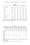
Purchased for the exclusive use of nofirst nolast (unknown)
From: SCC Media Library & Resource Center (library.scconline.org)