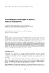
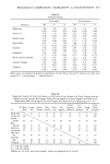
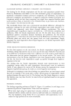
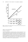
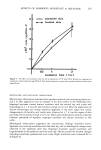
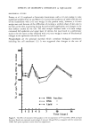
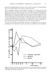
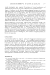
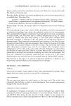
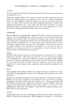
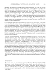
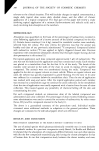
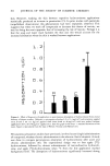
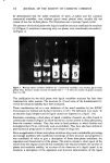
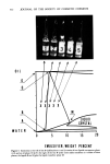
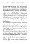
ANTIPERSPIRANT ACTION OF ALUMINUM SALTS 287 3+-- 2+-- 1+_ lOO 75 - 50 - 25 -- o i i i I I DAYS Figure 3. Type and intensity of millaria during anhidrosis induced by 24-hr occlusive exposure to 20% A1CI 3 ß 6H20. Severity of the lesions parallels sweat suppression decreasing gradually with time. Early on &l. rubra and &l. profun•ta predominate..84. crysta//ina becomes common after two weeks and is overwhelmingly dominant by three. aluminum anhidrosis could have been predicted. For the first week, the lesions were a mixture of 2•I. rubra and /14. profunda. By two weeks, 2•I. profunda was on the wane and 2•I. crysta//ina had become prominent. By three weeks, there was only one response, ./14. crystallina. This sequence is easily explained. The obstruction starts at a deep level and becomes progressively shallower, until it is located entirely within the horny layer. The early obstruction must extend into the dermis in order for the wheal-like 2•I. profunda lesions to develop. These resorb in a matter of minutes when sweating stops. A block within the intra-epidermal portion of the acrosyringium is the precondition for the development of 2•I. rubra. That both occur together in the early phase bears out what was already disclosed by the study of the duration of anhidrosis, viz., different ducts are blocked at different levels. It is logical, indeed inevitable, that the final lesion in the sequence be 2•I. crysta//ina, reflecting a superficial intra-corneal block. The progressive transformation of the initial deep block to an ever more superficial location is attributable to epidermopoiesis. It is appropriate to note here that a somewhat similar sequence of miliarial lesions was observed when anhidrosis was induced by occlusive patches for 3 to 4 days. This results in the formation of an obstructing agglomerate of degenerating leukocytes and PAS-positive masses within the lumen of the sweat ducts (8).

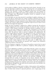
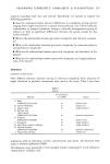
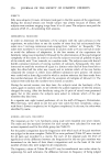
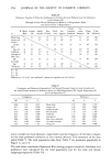
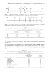
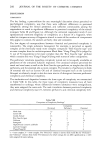
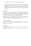
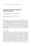
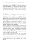
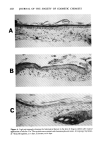
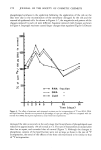
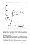
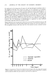
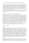
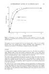
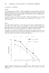
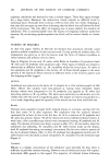
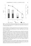
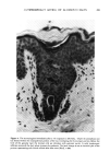
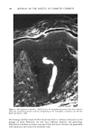
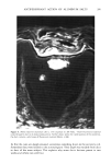
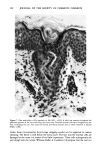
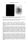
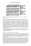
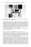
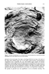
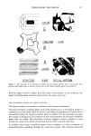
288 JOURNAL OF THE SOCIETY OF COSMETIC CHEMISTS HISTOLOGIC EXAMINATION The observations presented so far clearly implicate a physical block of some sort. But, with one exception, various observers have failed to visualize the obstruction. Relier & Luedders (14) found casts which took a reddish color with the aluminum stain. The cast was mainly within the "distal segment of the epidermal sweat duct," a finding somewhat incompatible with the long duration of the anhidrosis. However, they did occasionally find aluminum-staining material well below the epidermis. Method Anhidrosis was induced on the back of 16 subjects by either 3- or 24-hr occlusive patches of 20% AICI 3 ß 6H20. Full thickness specimens were excised shortly after the exposure or 7 days later. Prior to removal of the biopsies a short thermal sweat stimulus was given to verify that the site was anhidrotic. After formalin fixation serial 6 /am sections were cut and stained with H&E, PAS and the morin technique. With the latter, aluminum-containing material fluoresces brightly. Results After a 3-hour exposure and even more so after 24, an eosinophilic, amorphous cast was found within the lumen throughout the entire acrosyringium. The luminal cells of the intra-epidermal portion showed moderate toxic changes indicated by pyknotic nuclei and eosinophilic cytoplasm (Figure 4). The luminal masses were PAS-positive and diastase-resistant. The casts were vividly visualized by the fluorescent morin technique (Figure 5). Moreover, they could be shown to extend well into the dermal segments, sometimes occupying the ducts right down to the secretory coil. By contrast, the intra-corneal portion of the ducts was patent with only a thin layer of aluminum-containing material coating the keratinized cells (Figure 6). In all cases, a modest infiltrate of lymphocytes surrounded the ducts at about the level where these entered the epidermis. The histologic picture was consistent with a sub-clinical miliaria. We emphasize that this change was provoked by the thermal stress which preceded taking the biopsy. The periductal infiltrate did not occur when tissue was removed without prior sweating. Hence, inflammatory changes are not an intrinsic feature of aluminum anhidrosis. After a week, a somewhat different view greeted the eye. The damaged and now shrunken luminal c•11s had sloughed into the lumen, enveloping the amorphous material which was still strongly PAS-positive. This agglomerate extended throughout the acrosyringigum including now the stratum corneum (Figure 7). The mid-dermal ducts were patent. Occasionally, intra-corneal microvesicles typical of M. crystallina were observed. We note here in passing that aluminum chlorhydroxide, the partially neutralized salt of aluminum chloride, produced the same histologic changes but to a lesser degree. Comment Though most brilliantly visualized by the fluorescent morin technique, the casts were observed in both PAS and H&E stained specimens. It is utterly beyond our understanding how a brigade of investigators, including ourselves, had failed to see these.
Purchased for the exclusive use of nofirst nolast (unknown)
From: SCC Media Library & Resource Center (library.scconline.org)