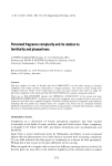
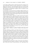
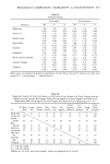
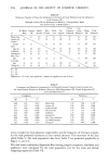
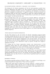
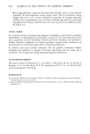
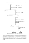
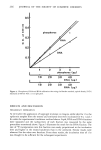
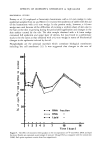
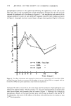
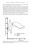
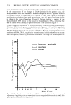
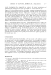
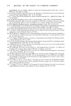
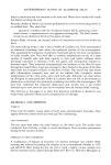
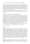
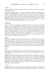
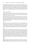
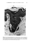
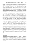
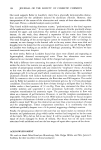
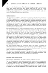
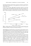
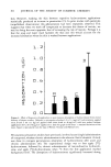
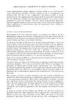
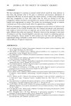
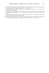
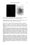
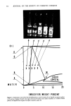
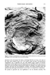
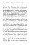
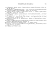
EFFECTS OF ISOPROPYL MYRISTATE & SQUALANE 265 skin samples incubation with 32p at 37øC for I hour extraction with CHCI3-CH3OH (2:1) lipid fraction resi I TCA-soluble fraction duo extraction with 5 % TCA residue 1 RNA fraction hydrolysis with I N KOH addition of 1! 10 portion of 70 95 HCIO 4 residue extract ion with I N HCff) 4 I I DNA fraction resi due Figure 1. Separation method for isolation of lipids, RNA and DNA. Standard solutions of RNA and DNA were prepared by dissolving yeast RNA and calf thymus DNA in 1 N KOH, respectively. A standard solution of phosphorus was prepared by dissolving purified KH2PO 4 in distilled water. Calibration curves were drawn between the concentration and the optical density by the colorimetric method described by Bartlett (10). As shown in Figure 2, linear relationship was ascertained in all cases.

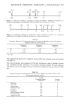
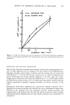
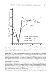
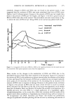
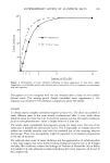
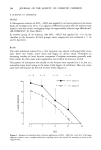
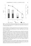
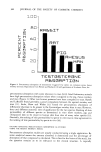
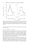
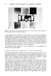
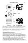
266 JOURNAL OF THE SOCIETY OF COSMETIC CHEMISTS 0., t E = 0.4 0.3 0.2 0.1 phosphorus • • DNA I I 10 20 I I 10 20 I I phosphorus (pg) 30 40 DNA (pg) I I , 30 40 RNA (pg) Figure 2. Phosphorus, DNA and RNA calibration data using the Bartlett method, optical density (O.D.) measured at 830 nm with a 1-cm light path. RESULTS AND DISCUSSSION PRELIMINARY EXPERIMENTS At 24 hr after the application of isopropyl myristate on Angora rabbit skin for 1 hr, the epidermis samples from the treated and untreated sites were incubated for 0.5, 1 and 2 hr under the experimental conditions outlined above. Lipid, RNA and DNA fractions were separated and the radioactivity of each fraction was measured by the same procedures mentioned above. Figure 3 illustrates the result for the DNA fraction. The rate of 32p incorporation into this fraction was nearly proportional to the incubation time and higher in the treated epidermis than in the untreated. Similar results were observed for the other two fractions. From these results, the incubation time of 1 hr was thought to be sufficient for the subsequent experiments.
Purchased for the exclusive use of nofirst nolast (unknown)
From: SCC Media Library & Resource Center (library.scconline.org)