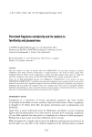
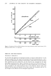
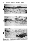
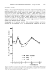
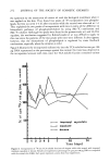
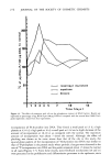
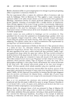
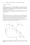
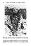
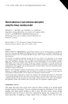
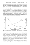
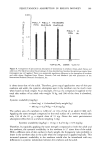
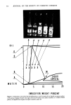
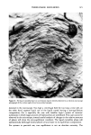
EFFECTS OF ISOPROPYL MYRISTATE & SQUALANE 269 BIOCHEMICAL STUDIES Prottey et al. (2) employed a Castroviejo keratotome with a 0.2-mm wedge to take epidermal samples from rat and Mezei (11) excised the epidermis of rabbit with the aid of the keratotome with a 0. l-ram wedge. In the present study, however, a 0.3-mm wedge was used because of the difficulties of excising a uniform sheet of skin due to the hair on the skin re-growing during the period after application and changes in the skin surface caused by the oils. The skin sample obtained with a 0.3-mm wedge contained full epidermis and upper layer of dermis, but was found in a preliminary study to be the same as that obtained with a 0.2-mm wedge in terms of biochemical changes in the epidermis induced by the oil. Phospholipids are the principal materials which constitute biological membranes including the cell membrane (12). It was suggested that changes in the rate of 200 ß 150B 100 50 ß ß RNA fraction ß , * DNA ,, ß ß Lipid ,, I I ! I, I, a I 2 3 4 5 7 ß ß ß _ 10 14 Time (days) Figure 5. The effect of treatment with squalane on the incorporations of 32p into RNA, DNA and lipid fractions. Results are expressed as percentage of cpm per 100 ttg DNA as compared with the normal skin (100%). Each point represents a mean from four experiments.

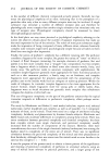
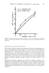
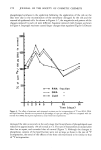
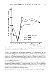
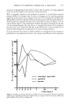
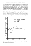
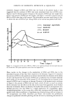
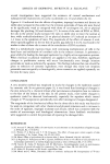
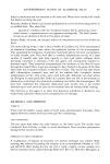
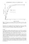
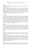
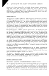
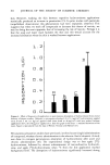
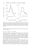
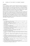
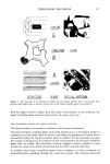
270 JOURNAL OF THE SOCIETY OF COSMETIC CHEMISTS phospholipid synthesis in the epidermis following the application of the oils on the skin were due to the reconstitution of the membrane damaged by the oils and the renewal of epidermal cells. As shown in Figures 5-7, the magnitude and pattern of the changes induced by each oil were different. Squalane induced mild changes as shown in Figure 5. Isopropyl myristate caused larger changes than squalane (Figure 6). Decane E200f o 150 100 50 m m m m m 1 ß ß RNA fraction ß ß DNA ,, ß ß Lipid ,, ß ß . II ß ß ! 2 3 4 5 7 10 14 Time (days) Figure 6. The effect of treatment with isopropyl myristate on the incorporations of •2p into RNA, DNA and lipid fractions. Results are expressed as percentage of cpm per 100/ag DNA as compared with the normal skin (100%). Each point represents a mean from four experiments. damaged the skin so severely in the early stage that biosynthesis of phospholipids was reduced to approximately 15% of normal at 3 d. The rate of biosynthesis increased after that due to repair, and exceeded that of normal (Figure 7). Although the changes in phosphorus content of the lipid fraction were not so large as those in the rate of 32p incorporation, the trend of the effects of the three oils was found to be similar to that of 32p incorporation.
Purchased for the exclusive use of nofirst nolast (unknown)
From: SCC Media Library & Resource Center (library.scconline.org)